
Open Access, Volume 11
The Leser‑Trélat sign as a marker of underlying malignancy
Anissa Lye Hui Min*
Senior Resident, Emergency Medicine, Tan Tock Seng Hospital, Singapore.
Anissa Lye Hui Min
Senior Resident, Emergency Medicine, Tan Tock Seng Hospital, Singapore.
Tel: +65-8182-9695;
Email: anissa.lye@gmail.com
Received : September 13, 2025,
Accepted : October 09, 2025
Published : October 31, 2025,
Archived : www.jclinmedcasereports.com
Copy right Statement: Content published in the journal follows Creative Commons Attribution License (http://creativecommons.org/licenses/by/4.0). © Min ALH (2025)
Journal: Open Journal of Clinical and Medical Case Reports is an international, open access, peer reviewed Journal mainly focused exclusively on the medical and clinical case reports.
Citation: Min ALH. The Leser‑Trélat sign as a marker of underlying malignancy. Open J Clin Med Case Rep. 2025; 2385.
Description
A 61-year-old Chinese lady with a background of a previous spinal cord tumour presented to the Emergency Department with severe pneumonia requiring ventilatory support. On examination, multiple eruptive seborrheic keratoses which had suddenly developed 3 years ago were incidentally noted over her back. Computed Tomography noted a uterine mass which correlated with a raised serum CA-125 level of 105, and a histology showed endometrial cancer. These findings were suggestive of the Leser-Trélat sign in association with a gynaecological malignancy.
The clinical photograph (Figure 1) depicts multiple warty, raised lesions with a ‘stuck-on’ appearance, on the patient’s back. She had a history of a previous spinal cord tumor. A serum CA-125 was raised, and a Computed Tomography scan found a suspicious-looking heterogenous 5.9 cm uterine mass corresponding histologically to endometrial cancer (Figures 2 & 3).
Leser-Trélat sign is a rare dermatological phenomenon characterized by sudden, rapid onset of multiple seborrheic keratoses, which are non-cancerous skin tumors. They occur in areas where seborrheic keratoses are not commonly found. It is a red flag for underlying malignancy, and anyone who exhibits this should seek medical evaluation.
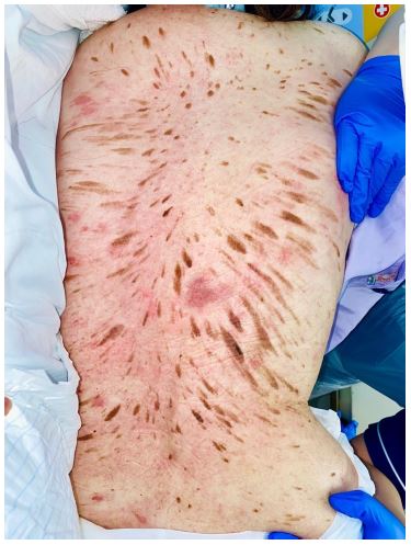
Figure 1: Leser–Trélat sign. Multiple seborrhoeic keratoses on patient’s back.
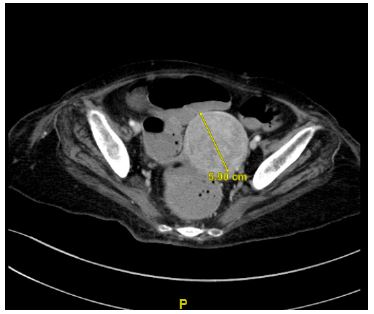
Figure 2: Axial CT of the patient’s pelvis showing 5.9 cm heterogenous uterine mass.
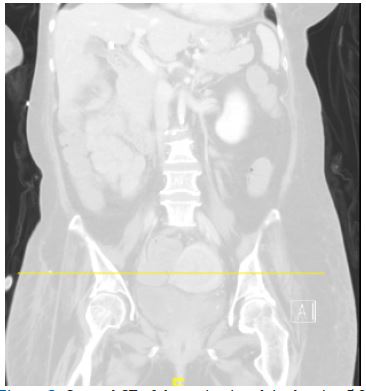
Figure 3: Coronal CT of the patient’s pelvis showing 5.9 cm heterogenous uterine mass.
Declarations
Conflict of interest statement: The author declares that they have no conflicts of interest related to this submission. No financial support or funding was received for this work.
