
Open Access, Volume 11
Laryngeal nodular fasciitis: A case report
Galea B*; Borg L; Correia E; Vale C; Muscat K
Department of Otorhinolaryngology, Mater Dei Hospital, Malta.
Galea B
Department of Otorhinolaryngology, Mater Dei Hospital, Malta.
Email: bernardgalea.7@gmail.com
Received : February 17, 2025,
Accepted : March 18, 2025
Published : March 28, 2025,
Archived : www.jclinmedcasereports.com
Abstract
Nodular fasciitis is a benign soft tissue tumour composed of myofibroblasts that typically develops in the extremities or trunk [3]. It often presents as a painless, firm and fast-growing mass in the subcutaneous tissue however it can also present with deeper areas of the limbs involving fascial layers [1]. It is often misdiagnosed with sarcoma due to its characteristically rapid growth [5,6]. It may be classified as a subtype of benign mesenchymal spindle cell tumours. Even though up to 25% of cases may present in the head and neck area, laryngeal nodular fasciitis is exceedingly rare [4]. In all 7 reported cases of laryngeal nodular fasciitis, patients presented with dysphonia or airway compromise [2,3].
Keywords: Nodular fasciitis; Dysphonia.
Copy right Statement: Content published in the journal follows Creative Commons Attribution License (http://creativecommons.org/licenses/by/4.0). © Galea B (2025)
Journal: Open Journal of Clinical and Medical Case Reports is an international, open access, peer reviewed Journal mainly focused exclusively on the medical and clinical case reports.
Citation: Galea B, Borg L, Correia E, Vale C, Muscat K. Laryngeal nodular fasciitis: A case report. Open J Clin Med Case Rep. 2025; 2336.
Case Presentation
We present the case of Mr. SA, an 84-year-old non-smoker, with a past medical history of controlled hypertension and type 2 diabetes, who presented with a 5-month history of intermittent dysphonia that became constant in nature over a period of 2 months. There was no history of GORD or recent endotracheal intubation.
Neck examination was unremarkable but flexible fiberoptic nasal endoscopy revealed a large exophytic mass originating from the middle third of the right vocal cord causing significant obstruction of the glottis (>80%). There was no evidence of stridor during the examination and the patient denied any shortness of breath. Both vocal cords were fully mobile, and no other mucosal lesions were visible.
An urgent CT neck with intravenous contrast was organized to further characterize the mass. Images revealed an exophytic polypoid lesion originating at the level of the glottis measuring 1.8(AP)×1.0(LL)×2.1(CC) cm (Figure 1). The lesion extended to occupy most of the airway causing significant narrowing of the airway itself but there was no evidence of infiltration of surrounding structures. There was no lymphadenopathy by CT size criteria in the neck.
The patient was scheduled for microlaryngoscopy and excision of the mass and was also consented for possible need of intraoperative tracheostomy, should airway access be difficult. Three days prior to the scheduled surgery the patient presented to clinic explaining that he had expectorated the mass spontaneously and that his dysphonia had resolved. Repeat flexible fiberoptic nasal endoscopy confirmed the absence of the mass with a visible pedicle origin over the anterior third of the right vocal cord and normal vocal cord mobility. The mass was sent for histological analysis. The case was discussed at our weekly ENT Multidisciplinary Team meeting.
Histology reported a polypoid ulcerated mucosal fragment, with proliferation of spindle cells, arranged in fascicles within an immature mucoid stroma. There are also scattered multinucleated giant cells noted, with a variable mixed inflammatory infiltrate within the stroma. Extravasated Erythrocytes are also seen. Immunohistochemistry showed a variable expression of alpha smooth muscle actin by the spindle cells, with no expression of desmin pancytokeratins (AE1/AE3), EMA, p63 or S100. CD68 highlighted the multinucleated giant cells as well as the stromal histiocytes with no evidence of malignancy.
The differential with this sort of picture includes rare laryngeal sarcomas, inflammatory myofibroblastic tumours, low grade myofibroblastic tumours and nodular fasciitis. The patient was followed up at outpatient clinic regularly. Two months following this episode of expectoration the patient developed hoarseness without stridor again. This time the presentation was induced by a lower respiratory tract infection. Repeat flexible fiberoptic nasal endoscopy revealed mass recurrence over the same area of the right vocal cord. The patient was scheduled for microlaryngoscopy and excision of the mass and was also consented for possible need of intraoperative tracheostomy, should airway access be difficult. The mass was removed successfully, and the root of the pedicle was cauterized with bipolar diathermy.
Histology reported a similar picture to original sample sent. The specimen reported as “ulcerated and inflamed granulation tissue focally covered by non-keratinised stratified squamous epithelium. Centrally there are areas of fasciculated spindle cell proliferation in a loose fibrous stroma with occasional keloidal collagen fibre bundles. Scattered mitotic figures are noted however there is no cytologic atypia and no other features of malignancy. Immunohistochemistry shows strong diffuse expression of á-smooth muscle actin and scattered expression of CD68 with weak equivocal expression of pancytokeratins (AE1/AE3) and no expression of keratins 5/6.”
Regular outpatients follow up confirmed no evidence of recurrence on flexible fiberoptic nasal endoscopy with significant improvement of dysphonia.
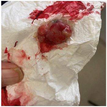
Figure 1: Specimen expectorated by the patient spontaneously.
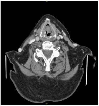
Figure 2: Contrast enhanced CT neck in axial view demonstrating lesion at the level of the glottis.
Discussion
The pathophysiology of nodular fasciitis is poorly understood and the exact etiology remains unknown [3]. The leading hypothesis describes the association of mechanical injury and a reactive inflammatory cascade, leading to over proliferation of myofibroblasts [4,6]. The patient described in this case report presented without direct injury or previous laryngeal exposure.
The literature reports dysphonia as the leading presenting complaint and other symptoms including dyspnoea and stridor with two cases reporting the need for emergency tracheostomy [2,7]. It is likely that most cases of laryngeal nodular fasciitis often go unreported. Size varies between the ranges of 0.5 to 10 cm, with the most common being less than 2 cm [3]. Rapid rate of growth and recurrence are commonly described in the literature [6].
Radiologically nodular fasciitis identifies as a well demarcated homogenous area of low to moderate enhancement on contrast enhanced computed tomography. It is reported to spread in different planes both radiologically and in terms of histocytology [3,6]. As per (Figure 2) below, we report a single-phase CT with the lesion showing minimal extension into the subglottic plane as well as cranial extension into the ventricle. There is no convincing evidence of deep extension into the paraglottic spaces laterally.
Treatment is typically that of surgical excision. Certain anatomical factors may render laryngeal nodular fasciitis lesions unamenable to microlaryngoscopy and excision and partial or total laryngectomy may be the only option in selected cases, particularly those larger than 2 cm [3,7]. Literature review also identifies the controversial use of intralesional corticosteroid injection when there are no substantial symptoms [6].
Conclusion
This case report highlights a rare presentation of nodular fasciitis. To our knowledge, there are 7 reported cases of laryngeal nodular fasciitis in the literature. The pathogenesis of these tumours is still unknown, but it is thought to be triggered by a local insult or inflammatory process that leads to proliferation of myofibroblasts. Laryngeal nodular fasciitis, unlike nodular fasciitis in other regions, is rarely asymptomatic and may compromise vocal cord function or present with airway compromise. In our case there was recurrence of growth of the mass following spontaneous expectoration. This point highlights the need of regular follow up with repeat fiberoptic nasal endoscopy after excision to assess for possible recurrence.
References
- Çelik SY, Dere Y, Çelik Öİ, Derin S, Şahan M, Dere Ö. Nodular fasciitis of the neck causing emergency: A case report. Oman Medical Journal. 2017; 32: 69–72.
- Nakagawa T, Sugimoto T, Komiyama S, Yamamoto T, Uemura T. Giant tumor formed by nodular fasciitis of the pharynx: a case report. Auris, Nasus, Larynx. 1994; 21: 196–199.
- Salunke A, Belgaumkar V, Chavan R, Dobariya R. Laryngeal involvement with fatal outcome in progressive nodular histiocytosis: A rare case report. Indian Dermatology Online Journal. 2016; 7: 516–519.
- Stadlhofer R, Böttcher A, Lübke A. First report of laryngeal nodular fasciitis. Deutsche Gesellschaft Für Hals-Nasen-Ohren-Heilkunde, Kopf- Und Hals-Chirurgie e.V., Bonn. 2021.
- Stadlhofer Rupert, Lübke A, Böttcher A. Laryngeal manifestation of nodular fasciitis: A case report and literature review. Cureus. 2021: 13.
- Svrakic M, Bent JP, Adler E. Neonatal nodular fasciitis of the larynx. International Journal of Pediatric Otorhinolaryngology. 2009; 73: 1007–1009.
- Völker HU, Scheich M, Höller S, Ströbel P, Hagen R, Müller-Hermelink HK, et al. Differential diagnosis of laryngeal spindle cell carcinoma and inflammatory myofibroblastic tumor--report of two cases with similar morphology. Diagnostic Pathology. 2007; 2: 1.
