
Open Access, Volume 11
Curation of non-healing cephalic wound treated with pulsed red LED light therapy after split skin transplantation
Magdalena Metzger1,2#; Leonie Krausgruber1,2#; Jonas Flatscher1,2; Peter Dungel1,2*
1Ludwig Boltzmann Institute for Traumatology, The Research Center in Cooperation with AUVA, Austria.
2Austrian Cluster for Tissue Regeneration, Austria.
#Equal Contribution.
Peter Dungel
Head of Photobiomodulation Research Group, Ludwig Boltzmann Institute for Traumatology, The
Research Center in Cooperation with AUVA, 1200 Vienna, Austria.
Email: peter.dungel@trauma.lbg.ac.at
#Equal Contribution
Received : January 10, 2025,
Accepted : February 06, 2025
Published : February 14, 2025,
Archived : www.jclinmedcasereports.com
Abstract
Basal Cell Carcinoma (BCC) is one of the most common human skin cancers and usually occurs on sun-exposed areas of the body. This carcinoma represents a public health concern due to its aggressive local invasiveness. Conventional treatment of neoplasms is based on surgical excision, but those interventions often result in extensive skin defects requiring autologous transplants like split thickness skin grafts. A possible negative consequence thereof is impaired blood circulation resulting in delayed- or non-healing wounds. A promising approach addressing this issue is Photobiomodulation (PBM), previously known as Low-Level Laser/Light Therapy (LLLT). Red or near infrared light, in particular, has been shown to enhance wound healing in both experimental models and humans.
We report here a 73-year-old Caucasian female patient with a non-healing wound high parietal on the scalp three years after split skin graft due to previous BCC excisions. The wound was treated with pulsed red 632 nm Light-Emitting Diode (LED) light (2.5 Hz, 50% duty cycle) twice a week for a period of three months with two breaks of two weeks each. The distance between the device and skin surface was 10 cm and the treatment time 9 min with a peak irradiance intensity of 100 mW/cm2, corresponding to a total light fluence of 27 J/cm2. During the irradiation, some sore areas healed while new inflammatory foci appeared, but complete healing was achieved roughly one month after the last therapy session. This concludes that LED light therapy might represent an effective therapeutic option for the management of postoperative non-healing wounds.
Keywords: Photobiomodulation; Basal cell carcinoma; Wound healing; Light therapy; Skin regeneration.
Abbreviations: BCC: Basal Cell Carcinoma; LED: Light-Emitting Diodes; LLLT: Low-Level Laser/Light Therapy; PBM: Photobiomodulation; VEGF: Vascular Endothelial Growth Factor.
Copy right Statement: Content published in the journal follows Creative Commons Attribution License (http://creativecommons.org/licenses/by/4.0). © Dungel P (2025)
Journal: Open Journal of Clinical and Medical Case Reports is an international, open access, peer reviewed Journal mainly focused exclusively on the medical and clinical case reports.
Citation: Metzger M, Krausgruber L, Flatscher J, Dungel P. Curation of non-healing cephalic wound treated with pulsed red LED light therapy after split skin transplantation. Open J Clin Med Case Rep. 2025; 2326.
Introduction
One of the most prevalent skin cancers in humans is Basal Cell Carcinoma (BCC) [1]. This epithelial skin cancer usually occurs on areas of the body which are heavily exposed to the sun, such as the neck or face. Although BCCs have a slow growth rate and low metastatic capacity, their tumors display high local invasiveness resulting in aggressive destruction of surrounding tissue [1,2]. Among the wide range of therapeutic modalities, surgical excisions are the most commonly applied based on their low recurrence rate [1,3].
Extensive skin cancer often requires the removal of large areas of skin, resulting in acute cutaneous wounds that demand further surgical interventions, e.g. autologous transplants [4,5]. After such transplantation, the graft has to undergo the steps for wound healing and one crucial process thereof is angiogenesis, which represents the development of new blood vessels out of a pre-existing vascular network [5]. Among other things, the new blood vessels are important for nutrient and oxygen delivery, indicating that defects in angiogenesis resulting in hypoxia and ischemia frequently lead to delayed wound healing [6]. Since skin grafts do not have their own blood supply, the underlying wound bed is responsible for their integration into the vascular network and thus, impairments thereof may result in complications of the split thickness skin graft [7]. One way to trigger angiogenesis is the usage of certain growth factors, for instance Vascular Endothelial Growth Factor (VEGF) [8].
Apart from that, several studies showed that light stimulation can also induce angiogenesis, hence, improve wound healing in general [9,11]. In short, Photobiomodulation (PBM), previously known as Low-Level Laser/Light Therapy (LLLT), uses lasers or Light-Emitting Diodes (LEDs) to minimize inflammation, relieve pain and generally aid in tissue regeneration [12]. This therapy is already known to be a suitable method with positive effects regarding wound healing in both experimental models and humans [13,14]. Especially red or near infrared light has been shown to promote injury repair in incisional wounds. This suggests that red light therapy may be suitable for postoperative wound management [15].
The aim of this case report is to show that low-level red LED light therapy represents an effective and non-invasive treatment for non-healing wounds after split-thickness skin grafting, even when they are located in areas where the skin is very tight and has little subcutaneous fat, such as the scalp.
Case Presentation
The case of a 73-year-old Caucasian female who was diagnosed with BCC in 2017 at the age of 67 is presented. Originally, two basaliomas were located high parietal (site A) and high occipital (site B) on the scalp which were surgically removed in September 2017. Two years later, additional excision procedures were necessary in July and August 2019 due to recurrence and failure to completely remove the BCCs.
After a total of five re-excisions, split skin extracted from the patient’s left thigh was grafted to the back of her head (site B) in August 2019. The transplant has healed very well but as the treatment was still not completely successful in terms of cancer removal, additional post-excisions were performed on January 14 and 21, 2020, resulting in a large open wound on the patient’s scalp (site A, Figure 1A). After the last excision, the BCC residues were successfully removed. Three days later, on January 24, 2020, a second split skin transplantation was performed to close the wound (site A). The skin graft was again removed from the left thigh of the patient.
During the entire treatment period, the patient was not prescribed any antibiotics and even despite this, she never had problems with wound infections. Wound care management after the surgical interventions involved standard procedures including compression bandages and frequent replacement of the bandage material. Furthermore, the patient covered her head with a wig or a scarf on a regular basis until the hair has grown back again on the surrounding healthy areas, meaning that the wound was not directly exposed to the environment during the healing process.
Since the graft (site A) had not healed completely even three years after the second split skin transplantation and a sore region was always present in the middle of the transplant area (Figure 1B), in September 2022, light therapy with red LED light was started to improve the healing process of the wound.
The transplanted skin on the head of the patient was treated 20 times over a period of three months with 2x2 weeks break in between, meaning that the wound was treated on average twice a week. Pulsed red LED light (Repuls 7, Repuls Lichtmedizintechnik GmbH, Vienna, Austria) with a wavelength of 632 nm, a pulse rate of 2.5 Hz and a duty cycle of 50% was used as a light source and applied for 9 min with 10 cm distance (Figure 1C) and a peak irradiance intensity of 100 mW/cm2 in each session, resulting in a light fluence of 27 J/cm2 (irridance was measured using the PM400K3 Optical Power Meter, Thorlabs, Bergkirchen, Germany). Besides the light treatment, the patient applied ‘Vitawund’ ointment (Haleon-Gebro Consumer Health GmbH, Fieberbrunn, Austria) and wore a wig on top of the wound during the days of therapy and afterwards.
Results and Discussion
An overview of the wound healing process during low-level LED light therapy is depicted in Figure 2. Within the first eight times of applying red light therapy, some regions were regenerated via crust formation and detachment, while new inflammatory foci appeared (Figure 2, first row). This pattern repeated itself between the 10th and 20th time of therapy. Overall, a reduction of the sore region was observed and after completion of the red-light therapy, the wounds were completely healed within about a month (Figure 2, last two images). A follow-up after approximately eight months showed a completely closed, non-inflamed transplant area (Figure 3), which has been maintained to date (two years later).
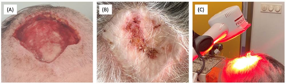
Figure 1: (A) Open wound after removal of basal cell carcinoma relapses (before split skin transplantation in January 2020). (B) Chronic wound at transplant area three years after split skin graft. (C) Treatment set-up of light therapy with pulsed red LED light (632 nm, 100 mW/cm2 peak, 27 J/cm2).
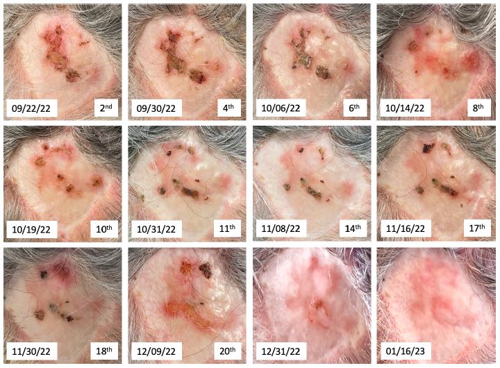
Figure 2: Course of the healing process of a scalp wound after split skin graft during light therapy with pulsed red LED light (632 nm, 100 mW/cm2 peak, 27 J/cm2). Images were taken directly before the respective treatment session. Treatment number and date are indicated at the bottom of each image. Last two images were taken three and five weeks after the final LED light therapy.

Figure 3: Split skin transplant area around eight months after completion of red LED light therapy.
Conclusion
In summary, we report a case of a non-healing graft area three years after split-thickness skin graft on the head of a 73-year-old female patient, which resulted in complete healing following treatment with 27 J/cm2 of 632 nm LED light. A follow-up of the graft area approximately two years later still showed complete closure, which could indicate that the light therapy successfully led to a permanently healed wound.
The used light source is a LED-based medical device that has already been applied in several in vitro studies. In particular, Dungel et al. observed positive results with this device regarding the stimulation of angiogenesis and wound healing in a diabetic mouse model [11]. Since the patient in the present case never had problems with wound infections, we assume that the issue was probably due to ischemia which may have been directly addressed with the light therapy.
In addition to the positive effects of PBM on wound healing, other benefits include cost-effectiveness, ease of application and the ability to use it as a home product. Therefore, red LED light offers a promising and effective treatment modality for wound care.
Acknowledgements: This research was partially funded by the AUVA Research Grant No. FK21/22 Phototherapy.
References
- K Tanese. Diagnosis and Management of Basal Cell Carcinoma. Curr Treat Options in Oncol. 2019: 20.
- KK Youssef, et al. Identification of the cell lineage at the origin of basal cell carcinoma. Nat Cell Biol. 2010; 12: 299–305.
- JYS Kim, et al. Guidelines of care for the management of basal cell carcinoma. J Am Acad Dermatol. 2018; 78: 540–559.
- MG Tonnesen, X Feng, RAF Clark. Angiogenesis in Wound Healing. J Investig Dermatol Symp Proc. 2000; 5: 40–46.
- DS Masson‐Meyers, L Tayebi. Vascularization strategies in tissue engineering approaches for soft tissue repair. J Tissue Eng Regen Med. 2021; 15: 747–762.
- KE Johnson, TA Wilgus. Vascular Endothelial Growth Factor and Angiogenesis in the Regulation of Cutaneous Wound Repair. Advances in Wound Care. 2014; 3: 647–661.
- ME Braza, MP Fahrenkopf. Split-Thickness Skin Grafts. in StatPearls, Treasure Island (FL): StatPearls Publishing. 2023.
- W Michlits, R Mittermayr, R Schäfer, H Redl, S Aharinejad. Fibrin‐embedded administration of VEGF plasmid enhances skin flap survival. Wound Repair Regeneration. 2007; 15: 360–367.
- IS Park, PS Chung, JC Ahn. Adipose-derived stromal cell cluster with light therapy enhance angiogenesis and skin wound healing in mice. Biochem Biophys Res Commun. 2015; 462: 171–177.
- S Rohringer, et al. The impact of wavelengths of LED light-therapy on endothelial cells. Sci Rep. 2017; 7: 10700.
- P Dungel, et al. Low level light therapy by LED of different wavelength induces angiogenesis and improves ischemic wound healing: LOW LEVEL LIGHT THERAPY BY LED. Lasers Surg Med. 2014; 46: 773–780.
- P Avci, A Gupta, M Sadasivam, Z Pam, N Pam, MR Hamblin. Low-level laser (light) therapy (LLLT) in skin: stimulating, healing, restoring. Semin Cutan Med Surg. 2013; 32: 41–52.
- RC Mosca, AA Ong, O Albasha, K Bass, P Arany. Photobiomodulation Therapy for Wound Care: A Potent, Noninvasive, Photoceutical Approach. Adv Skin Wound Care. 2019; 32: 157–167.
- FLC Pereira, MVL Ferreira, P Da Silva Mendes, FM Rossi, MP Alves, et al. Use of a High-Power Laser for Wound Healing: A Case Report. J Lasers Med Sci. 2020; 11: 112–114.
- Erdle, S Brouxhon, M Kaplan, J Vanbuskirk, AP Pentland. Effects of Continuous-Wave (670-nm) Red Light on Wound Healing. Dermatol Surg. 2008; 34: 320–325.
