
Open Access, Volume 11
Carpal tunnel syndrome secondary to collagenase treatment for Dupuytren’s disease
Ariadny Lobo-Alcala1,2; Rafael Sanjuan-Cervero1,3*
1Departament of Orthopedics and Trauma Surgery, Hospital de Denia, Alicante, Spain.
2Doctoral student, University of Valencia, Spain
3PhD. University of Valencia, Spain.
Rafael Sanjuan-Cervero
Department of Orthopedics and Trauma Surgery, Hospital de Denia, Alicante, Spain.
Email: sanjuan.rafcer@gmail.com
Received : January 05, 2025,
Accepted : February 04, 2025
Published : February 14, 2025,
Archived : www.jclinmedcasereports.com
Abstract
Injection of collagenase from Clostridium Histolyticum (CCH) in Dupuytren’s disease weakens diseased cords sufficiently to enable subsequent rupture and finger extension. The effects of CCH, however, are limited to the injection area, meaning that palpable traces of cord remain proximally and distally to the injection site. CCH weakens cords by breaking up collagen fibers, with residual fragments being subsequently degraded by post-treatment inflammatory responses. Involvement of the septa of Legueu and Juvara in Dupuytren´s disease could give rise to a mechanical phenomenon whereby a tunnel would be formed around the course of the median nerve and tendon sheaths. The local action of CCH would dissolve just the roof of the tunnel, leaving the other areas intact and entrapping a nerve. We present the case of a patient with a middle finger Metacarpophalangeal (MCP) joint contracture treated with CCH in whom residual cord proximal to the injection site caused compression of the median nerve near the transverse carpal ligament in the palm of the hand. Carpal tunnel is a rare adverse effect that can occur in patients with long, thick cords pretendinous cords treated with local CCH injection.
Keywords: Collagnease clostridium histolyticum; Carpal tunnel; Dupuytren´s diasease; Fasciectomy; Median nerve.
Abbreviations: CCH: Collagnease Clostridium Histolyticum; MCP: Metacarpo-Phalangeal Joint; EMG: Electromyography; CTS: Carpal Tunel Syndrome; URAM: Unité Rhumatologique des Affections de la Main scale.
Copy right Statement: Content published in the journal follows Creative Commons Attribution License (http://creativecommons.org/licenses/by/4.0). © Sanjuan-Cervero R (2025)
Journal: Open Journal of Clinical and Medical Case Reports is an international, open access, peer reviewed Journal mainly focused exclusively on the medical and clinical case reports.
Citation: Lobo-Alcala A, Sanjuan-Cervero R. Carpal tunnel syndrome secondary to collagenase treatment for Dupuytren’s disease. Open J Clin Med Case Rep. 2025; 2325.
Introduction
IInjectable Collagenase Clostridium Histolyticum (CCH) is a nonsurgical alternative for the treatment of Dupuytren disease. It is less aggressive than fasciectomy and offers significantly faster recovery times [1]. While adverse effects are very common, they are mostly mild and self-limiting [2]. Severe adverse effects are rare and include tendon rupture, persistent pain, complex regional pain syndrome, and anaphylactic reactions [3,4]. Skin tear is perhaps the most significant adverse effect for patients [5]. CCH weakens pathologic cords by breaking up the collagen fibers, with residual fragments being subsequently degraded by post-treatment inflammatory responses [6-9]. Treated cords are often no longer palpable after finger manipulation (generally performed the next day), and in some cases, the treated finger can be extended spontaneously. The effects of CCH injection, however, are limited to the area around the injection site, meaning that palpable traces of cord remain proximal and distal to this site. While residual cord does not generally cause problems, several authors recommend distributing the entire contents of the vial along the cord [10] or using several spots rather than changing the direction of the needle [11]. Although residual cords are typically an aesthetic concern, their persistence can be problematic in cases.
We present the case of a patient with a middle finger Metacarpophalangeal (MCP) joint contracture treated with CCH who developed carpal tunnel syndrome shortly after treatment, possibly due to compression of the median nerve by residual cord proximal in the palm. The patient provided informed consent for the publication of his case.
Case Report
59-year-old man with a personal history of severe alcoholism and epilepsy presented with a 48° MCP joint contracture and a 28° proximal interphalangeal joint contracture in the ring finger. The cord also extended in a Y towards the radial area of the middle finger (Figure 1). The MCP joint was injected with 0.58 mg of CCH, and the remaining volume of the vial was injected into the middle finger (one spot in the area of the radial cord on the palm and another proximally to the distal palmar crease). The patient showed improvement at the 1-month follow-up, with a ring finger contracture of just 12 degrees (Figure 2) and full extension of the middle finger. There were palpable traces of cord proximal to the distal palmar crease. No clinical or functional problems were reported (score of 0 on the Unité Rhumatologique des Affections de la Main scale). months after treatment, the patient started to show signs of median nerve compression in the carpal tunnel characterized by tingling and numbness (mostly at nighttime) in the area of the three radial digits. He also experienced a loss of fine touch sensation (inability to pick up coins, button up a shirt, or hold a telephone during a call). The presence of residual cord proximal to the palm meant it was not possible to test for the Tinnel sign. The Phalen sign was suggestive of median nerve compression. Electromyography (EMG) revealed mild, predominantly sensory, mononeuropathy of the median nerve. Integration of clinical and EMG findings led to a diagnosis of Carpal Runnel Syndrome (CTS). On considering the relationship between the effects of CCH treatment, the patient’s renewed ability to fully extend his finger, and the immediate onset of CTS symptoms, we suspected that the palpable cord detected may have had a causative role. Surgery was performed four months after diagnosis and consisted of subcutaneous resection of the palmar cord from the distal end of the transverse carpal ligament to the distal palmar crease followed by open surgical release of the transverse carpal ligament (Figures 3-5). The patient showed progressive improvement and had achieved full functional recovery by the 4-month follow-up visit.
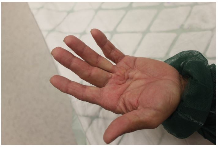
Figure 1: Dupuytren´s disease before treatment.
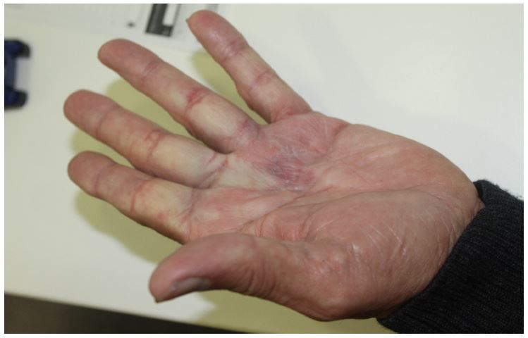
Figure 2: Hand after treatment. One month follow up.
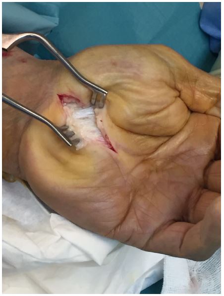
Figure 3: Diffuse affectation of the palmar fascia proximal to the proximal palmar crease.
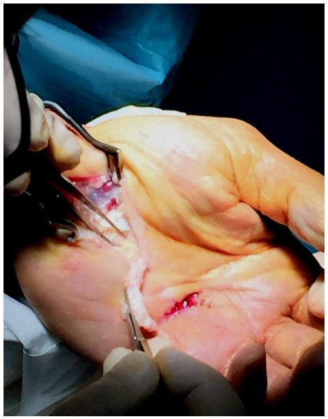
Figure 4: Partial proximal fasciectomy and subcutaneous release of the palmar cord of the ring finger.

Figure 5: Carpal tunnel decompression.
Discussion
To our knowledge, there has been just 1 other report of CTS after CCH injection, and it also involved the middle finger [12]. We agree with the authors of this initial report that CCH was unlikely to be responsible for the CTS observed, as the product was injected at a significant distance from the carpal tunnel. Unlike the authors, however, we do not think that the nerve compression was caused by post-treatment swelling. Rather, we believe that it was the result of a mechanical phenomenon linked to the presence of a thick residual cord in the proximal palm and involvement of the septa of Legueu and Juvara. Combined, these would create a tunnel (whose roof would correspond to the cord and whose sides would correspond to the septa). The action of CCH would only destroy the roof, leaving the sides intact. The septa of Legueu and Juvara connect the palmar aponeurosis to the deep structures of the palm. In this particular case, they could create a mechanical phenomenon whereby the median nerve and tendon sheaths would be partially released around the injection site, but remain compressed by the undissolved Dupuytren cord in more proximal areas. In other words, in cases with involvement of the septa of Legueu and Juvara and proximal palmar extension, administration of CCH at the level of the MCP could cause a contradictory effect by which the nerve and tendon would be released around the injection site but compressed by residual cord proximal to the distal palmar crease. In our patient, we believe that this process caused a bowstring-like effect characterized by a narrow area proximal to the injection site formed by the end of the carpal tunnel and deep expansions of residual cord in the proximal palm. Likewise, finger extension would have caused compression in the area around the end of the residual cord when the patient regained full extension of his hand. This case was resolved by resecting the residual cord and releasing the carpal tunnel. Similarly to in the treatment of Dupuytren’s disease, release of the proximal cord via subcutaneous surgery decompressed the nerve, making it unnecessary to completely resect the cord at the septal level. It may not have been necessary to open the carpal tunnel, but in light of the clinical diagnosis, we decided that it would be a safer option.
When treating Dupuytren disease with predominant MCP involvement, we generally inject 0.58 mg of CCH (the dose specified in the Summary of Product Characteristics) into the affected joint and then administer the remaining volume proximally or distally to minimize the risk of recurrence and improve aesthetic outcomes [10]. Administration of CCH along a greater length of cord reduces subsequent problems and creates a stronger visual effect of the cord having “disappeared”. It also improves patient satisfaction, as patients achieve better finger extension or even hyperextension after treatment, although these results have not been quantified. Administration of a higher-than-indicated dose could obviously increase the risk of adverse effects, but these are mild in the majority of cases [4].
The rapid onset of CTS symptoms led us to suspect a secondary cause. Although EMG showed mild median neuropathy in our patient, clinical manifestations of CTS often do not correlate with EMG findings [13]. The manifestations in the present case, however, indicated probable CTS [14], and this suspicion was confirmed by the improvement observed after surgery. The main limitation of this case report is that we did not use imaging tests to confirm the diagnosis of CTS or administer tests (e.g., URAM scale [15] or Levine Questionnaire [16]) to assess severity and functional status before and after treatment.
Conclusion
In brief, we believe that our patient developed CTS as the result of a mechanical phenomenon caused by the presence of a thick cord extending proximally to the palmar crease with involvement of the septa of Legueu and Juvara, leading to the formation of palmar tunnels separating the tendon and neurovascular structures. The action of the CCH would have eliminated the roof of the tunnel, creating a compression area adjacent to the injection site.
References
- Inês M, Silverio NM, Erdogan-Ciftci E. PMS43 Cost-Minimization Analysis of Collagenase Clostridium Histolyticum Compared with Fasciectomy in Patients with Dupuytren’S Contracture in Portugal. Value in Health. 2011; 14: A310.
- Hurst LC, Badalamente MA, Hentz VR, et al. Injectable Collagenase Clostridium Histolyticum for Dupuytren’s Contracture. New England Journal of Medicine. 2009; 361: 968-979.
- Peimer CA, Wilbrand S, Gerber RA, Chapman D, Szczypa PP. Safety and tolerability of collagenase Clostridium histolyticum and fasciectomy for Dupuytren’s contracture. J Hand Surg Eur Vol. 2015; 40: 141-149.
- Sanjuan-Cerveró R, Carrera-Hueso FJ, Vazquez-Ferreiro P, Gomez-Herrero D. Adverse Effects of Collagenase in the Treatment of Dupuytren Disease: A Systematic Review. BioDrugs. 2017; 31: 105-115.
- Sanjuan-Cervero R, Carrera-Hueso FJ, Oliver-Mengual S, Ramon-Barrios MA, Peimer CA, Fikri-Benbrahim N. Skin Laceration in Collagenase Clostridium histolyticum Treatment for Dupuytren’s Contracture. Orthop Nurs. 2018; 37: 144-153.
- Bond MD, Van Wart HE. Purification and separation of individual collagenases of Clostridium histolyticum using red dye ligand chromatography. Biochemistry. 1984; 23: 3077-3085.
- Bond MD, Van Wart HE. Characterization of the individual collagenases from Clostridium histolyticum. Biochemistry. 1984; 23: 3085-3091.
- Bond MD, Van Wart HE. Relationship between the individual collagenases of Clostridium histolyticum: evidence for evolution by gene duplication. Biochemistry. 1984; 23: 3092-3099.
- Radice M, Brun P, Bernardi D, Fontana C, Cortivo R, Abatangelo G. Clostridial collagenase releases bioactive fragments from extracellular matrix molecules. J Burn Care Rehabil. 1999; 20: 282-291.
- Atroshi I, Nordenskjöld J, Lauritzson A, Ahlgren E, Waldau J, Waldén M. Collagenase treatment of Dupuytren’s contracture using a modified injection method. Acta Orthop. 2015; 86: 310-315.
- Warwick DJ, Graham D, Worsley P. New insights into the immediate outcome of collagenase injections for Dupuytren’s contracture. J Hand Surg Eur Vol. 2015.
- Shubinets V, Lin IC, Chang B. Carpal Tunnel Syndrome after Xiaflex Injection for Dupuytren Disease. Plast Reconstr Surg. 2017; 139: 1031e-1032e.
- Chan L, Turner JA, Comstock BA, et al. The relationship between electrodiagnostic findings and patient symptoms and function in carpal tunnel syndrome. Arch Phys Med Rehabil. 2007; 88: 19-24.
- Rempel D, Evanoff B, Amadio PC, et al. Consensus criteria for the classification of carpal tunnel syndrome in epidemiologic studies. Am J Public Health. 1998; 88: 1447-1451.
- Gómez-Herrero D, Carrera-Hueso FJ, Sanjuan-Cerveró R, et al. Validation of a spanish version of the ‘Unité Rhumatologique Des Affections De La Main’ (URAM) scale. Journal of Plastic, Reconstructive & Aesthetic Surgery. 2021; 74: 1621-1628.
- Levine DW, Simmons BP, Koris MJ, et al. A self-administered questionnaire for the assessment of severity of symptoms and functional status in carpal tunnel syndrome. J Bone Joint Surg Am. 1993; 75: 1585-1592.
