
Open Access, Volume 11
A comprehensive management of cytostatics extravasation lesions: An overview
Dayamí Zaldívar Castillo, MD1*; Sady Valdés Mesa, MD2; Clara Elena Peñalver Rodríguez3; Juan Camilo Zafra Henao, MD4; Javier Ricardo Meriño, MD4
1Specialist 2nd Degree, Plastic Surgery, Department of Burns and Reconstructive Surgery, General Calixto García University Hospital, Cuba.
2Specialist 2nd Degree, Plastic Surgery, Department of Burns and Reconstructive Surgery, Luis Díaz Soto Hospital, Cuba.
3Teaching Specialist, Department of Teaching and Research, General Calixto García University Hospital, Cuba.
4Resident, Plastic Surgery, Department of Burns and Reconstructive Surgery, General Calixto García University Hospital, Cuba.
Dayami Zaldívar Castillo
Specialist 2nd Degree, Plastic Surgery, Department of Burns and Reconstructive Surgery, General Calixto García University Hospital, Havana, Cuba.
Email: mbctellez@gmail.com
Received : January 01, 2025,
Accepted : February 03, 2025
Published : February 14, 2025,
Archived : www.jclinmedcasereports.com
Abstract
Introduction: Cytostatics are antineoplastic drugs of vital importance and can be administered parenterally via a central or peripheral intravenous route. Despite advances in therapeutic techniques, the extravasation of chemotherapy drugs continues to be a fatal accident that leads to tissue necrosis, with a decrease in the quality of life of those affected and the need for early diagnosis and effective therapy, including surgery. With the aim of describing the state of the art of this complication, as well as the different therapeutic criteria, this review is carried out.
Methods: A systematic review of the medical literature on articles referring to cytostatic extravasation was carried out from January 2023 to August 2024. The databases used were: Pubmed, Infomed, Scielo, academic Google.
Results: 35 articles related to cytostatic extravasation were included.
Discussion: The inadvertent escape of cytostatic drugs from the blood vessels into the subcutaneous cellular tissue constitutes an unwanted and stressful complication, sometimes undiagnosed, and can cause irreversible damage to surrounding tissues, including necrosis and functional impotence of the affected limb.
Conclusions: Early detection and a comprehensive, standardized and interdisciplinary therapeutic approach must be implemented to reduce the negative consequences of lesions due to cytostatic extravasation.
Keywords: Cytostatics; Extravasation; Therapeutics; Necrosis.
Copy right Statement: Content published in the journal follows Creative Commons Attribution License (http://creativecommons.org/licenses/by/4.0). © Castillo DZ (2025)
Journal: Open Journal of Clinical and Medical Case Reports is an international, open access, peer reviewed Journal mainly focused exclusively on the medical and clinical case reports.
Citation: Castillo DZ, Mesa SV, Rodriguez CEP, Henao JCZ, Merino JR. A comprehensive management of cytostatics extravasation lesions: An overview. Open J Clin Med Case Rep. 2025; 2324.
Introduction
Cytostatics are a group of drugs of vital importance for the treatment of cancer, because of its capacities of inhibit the growing of tumoral cells [1]. Generally they are administered intravenously, using central s or peripheral access [2]. In spite of the advances in therapeutics thecniques, they can cause adverses reactions, including the extravasation or escape of chemotherapics from a blood vessels during the infussion, damaging the surrounding tissues [3]. The severity of the accident depends on the quantity and quality of extravasated medication [2].
Peripheral veins are located in the subcutaneous celular tissue, a laxus fatty space, facilitating an easily collection of extravasated chemotherapics, so a damage of surrounding tissues occurs, involving skin, nerves, vessels, tendons and muscles [3,4].
The citotoxic properties of the drugs causing lesions of the tissues are the results of two main action mechanisms: (1) Absortion of the drugs by local cells, binding the microtubules and the celular DNA, and number, (2) Direct damage of tissues without affecting the DNA [4,5].
Citostatics extravasation lesions are considered a medical urgency, because of the severity of complications developed, including the temporary interruption of the specific oncological treatment, and affecting the quality of patient´s life [6].
Regarding the accidental nature of this complication, there are not specifics statisticals reports about the incidence of chemotherapy extravasation [7,8].
A proper knowledge of the pathophysiology of cytostatics extravasation lesions is important in order to determinate an early diagnosis and for an integral therapeutic approach, including a conservative treatment or a surgery, with the debridement of the necrotic tissues and a definitive coverage of the wound [9]. With the aim to describe the status of the art of cytostatics extravasation events, and the differents treatment criterias ,a review of the medical literature was carried out.
Methods
A systematic review of the medical literature was carried out from January 2023 to August 2024. The searched databases included: Pubmed, Infomed, Scielo, academic Google. Medical Subject Headings (MeSH) were used for searching: Cytostatics Agents, extravasation, therapeutics, necrosis.
Medical articles related to chemotherapy extravasation lesions published from the year 2000 onwards, were searched.
Inclusion and Exclusion Criteria
− Inclusion criteria: Originals articles, Case presentations and review articles related to cytostatics extravasation.
− Exclusion Criteria:
Medical articles related to extravasation events, differentes to cytostatics medication
Articles published before the year 2000.
Results
Of a total of 92 medical articles searched, 35th were included, distributed as follows: 21 reviews, 9 original articles and 5 case reports. Of them, 12 articles were published during the last 5 years (2019 to 2024).
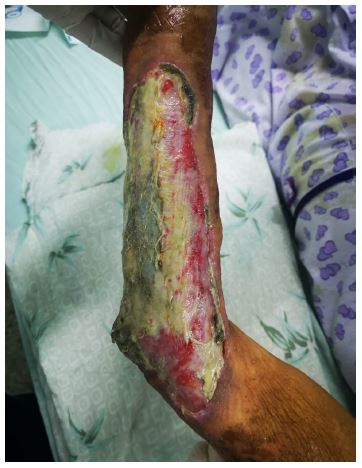
Figure 1: Cytostatic extravasation of the forearm.
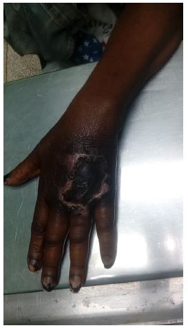
Figure 2: Cytostatic extravasation of the hand.
Discussion
The inadvertent leakage of cytostatics drugs from a blood vessels to extravascular space during intravenous administration, is a stressfull and undesired complication, sometimes undiagnosed, and can results in severe and irreversibles tissue damages, including necrosis, and functional impairment of the affected extremity [12,13].
Several risks factors appears to be related to the development of this complication. García et al, [14] report local factors as venous fragility and sclerosis, previous irradiation of the injection site, presence of linfedemas, multiples venopunctures. Others general factors as: extremes ages (ancients or children), immunodepressions conditions, malnutrition. somnolence status [15,16].
Some predisposing factors depending on the technique are described: a proper selection of the size and type of intravenous cannula, selection of an optimal peripheral vein and site of injection, avoiding multiples punctions as well as the use of infussion pumps and butterflys [17,18]. A right competence and experience of the paramedical and medical team is required, to avoid an extravasation events. Prevention suposed to be the first line of attention [19,20].
An adequate education of patient for an early recognition of symptoms as: redness, pain, itching, burning, swelling, is essential for the instauration of an early treatment [21,22].
An inmediate intervention, interrupting the infussion of the medication, and remotion of the intravenous cannula is vital [22-24].
Vasoconstrictive properties of cold compresses applied to diminish the velocity of drug difussion in the subcutaneous space, and acute inflammation signs have been reported [24]. The usefullness of topic hot to increase the absortion of the extravasated cytostatic drug is based on its vasodilatation effects [24-26].
There is a lack of consensus regarding the Classification of antineoplastic agents [27]. According to their citotoxic capacities, cytostatics agents are classified in: Non agresives, Irritants, and Vesicants [22,25,27]. The vesicants ones are responsibles for necrotic and deep tissue injuries. Upper extremities are the most used anatomical areas for peripheral intravenous drug administration [28,29].
Antecubital fose have been signaled as the first site affected for extravasation events [30]. Alfaro Rubio [9] reports the dorsus of the hand as the main affected anatomic zone, (Figure 1) followed by the forearm (Figure 2). The dorsal zone of the foot results the most affected site injection in children [28,31].
Pluschnig [23] have preconized the usefullness of contrasted angiography with green indocianine for the evaluation of blood circulation in the extravasated area, it confirms the extensión of the damaged tissue, and helps to decide whether to apply a surgical or conservative treatment.
Pérez Fidalgo et al, [21] confirm the effectiveness of topic application of DMSO (dimetylsulfoxide 50%), inmediately after the extravasation, and twice a day for two weeks. Its a common solvent, that penetrates tissues, increasing the velocity of elimination of extravasated drug.
Enzymatic debridement of necrotic tissue with hialorunidasa have been described by several authors [31].
The effectiveness of topical steroids have not been confirmed [32]. Darmert et al, [33] inform that the topical use of antidotes as Dexrazoxane is considered controversial and its not an standard procedure.
The surgical debridement and the subsequent surgical coverage using local flaps or skin grafts have been demostrated to be the definitive treatment [34], that is why the early reference of affected patients to the Plastic Department for surgery, is advisible [35].
Jaime Fagundo et al [6] consider the amputation surgery as an exceptional approach, when the severity of the injury results in an impairment extremity viability [35].
The role of the early hyperbaric oxigenation therapy have been reported [33].
Conclusions
An early diagnosis and an integral standarized therapeutic approach most be implemented to diminish the negative effcts of cytostatics extravasation lesions.
Conflict of interest: No conflicts of interests are declared between the authors.
References
- Jackson Rose J, Del Monte J, Groman A, Dial LS, Atwell L, Graham J, Rice RD. Chemotherapy extravasation: Establishing a national benchmark for incidence among cancer centers. Clinical Journal of Oncology Nursing. 2017; 21: 438-455.
- Gallieni M, Pittiruti M, Biffi R. Vascular Access in Oncology patients. CA: A Cancer Journal for Clinicians. 2008; 58: 323-346.
- Ramos PL. Efectos adversos dermatológicos a agentes quimioterápicos. Rev Col Hematol Oncol. 2022; 9: 77-79.
- Barbee MS, Owonikoko TK, Harvey RD. Taxanes: vesicants, irritants, or just irritating?. The Adv Med Oncol. 2014; 6: 16-20.
- Batista Lima SFB, Díaz Fernández da Conceicao MI, de Sá Tinoco JD. Diagnostic accuracy of the Clinical indicators of vascular trauma in patients undergoing antineoplastic chemotherapy in peripheral veins. Journal of Vascular Nursing. 2023; 41: 149-152.
- Jaime Fagundo JC, Arencibia Núñez A, Romero González A, Anoceto Martínez A, Pavón Morán V. Urgencias en Hematología II: Extravasación de citostáticos. Rev Cub Hematología, Inmunología y Hemoterapia. 2012; 28: 120-129.
- Ehmke N. Chemotherapy Extravasation: Incidence of and factors Associated with events in a Community Cancer Center. CJON. 2021; 25: 680-686.
- Moyle P, Soh Ch, Healy N, Malata Ch, Forouchi P. Extravasation of Epirubicin chemotherapy from a port-a-cath causing extensive breast necrosis: Sequential imaging findings and management of a breast cancer patient. Radiology Case Reports. 2021: 3509-3514.
- Alfaro Rubio A, Sanmartín O, Requena C, Llombart B, Botella Estrada R, Nagore E, Serra Guillén C, Hueso L, Guillén C. Extravasación de agentes citostáticos: una complicación grave del tratamiento oncológico. Actas Dermosifiliogr. 2006; 97: 169-176.
- Nguyen M, Borders L, Wesolow JT, Greene J. Chemotherapy Extravasation Causing Soft-Tissue Necrosis Mimmicking Infection: A Longitudinal Case Study. Cureus. 2024; 16: e55333.
- Pham TD, Tsunoyama T. Exploring Extravasation in Cancer Patients. Cancers. 2024; 16: 2308.
- Boschi R, Rostagno E. Extravasation of antineoplastic agents: prevention and treatment. Pediatrics Reports. 2012; 4: e28.
- Hassan DS, Hasary HJ. Chemotherapy-Induced Extravasation Injury: Classification and Management. Al-Rafidain J Med Sci. 2022; 2: 81-92.
- García Sánchez D, Santa Cruz ME, Chongo Solís C. Prevención y tratamiento de de la extravasación de quimioterapia intravenosa. Rev Cubana de Enfermería. 2019; 35: e1889.
- Kriedieh FY, Moukadem HA, El Shaghir NS. Overview, prevention and management of Chemotherapy extravasation. World J Clin Oncol. 2016; 7: 87-97.
- Antúnez Blancat A, Gago Valiente FJ, García Iglesias JJ, Merino Navarro D. The Role of Nursing in the Management of Chemotherapy Extravasation: A Systematic Review Regarding Public Health.Healthcare. 2024; 12: 1456.
- Kim SS, Holcombe RF. Development of chemotherapy –induced extravasation management algorithm to facilitate and improve care of cancer patients. Journal of Clinical Oncology. 2014; 32.
- Hale O, Deutsh PG, Lahiri A. Epirubicin extravasation: consequences of delayed management. BMJ Case Rep. 2017: 1-3: bcr2016218012.
- Langstein HN, Duman H, Seelig D, Butler Ch E, Evans GR. Retrospective Study of the Management of Chemotherapeutic Extravasation Injury. Annals of Plastic Surgery. 2002; 49: 369-374.
- Karius DL, Colvin CM. Managing Chemotherapy Extravasation Across Transitions of care: A Clinical Nurse Specialist Initiative. Journal of Infusion Nursing. 2021; 44: 14-20.
- Pérez Fidalgo JA, García Fabregat L, Cervantes A, Margulies A, Vidall C, Roila F. Management of Chemotherapy extravasation: ESMO- EONS Clinical Practice Guidelines. Annals of Oncology. 2012; 23. vii167-vii173.
- Karczmarek Borowska B, Matczuk M. Management of extravasation of drugs applied in cancer therapy. Contemporary Oncology. 2004; 8: 29-32.
- Pluschnig U, Haslik W, Bartsch R, Mader RM. Extravasation emergencies: state –of-the-art management and progress in Clinical Research.memo. 2016; 9: 226-230.
- Goolsby TV, Lombardo FA. Extravasation of Chemotherapeutic Agents: Prevention and Treatment. Seminars in Oncology. 2006; 33: 139-143.
- Al SA, Asha W, Abou Char MK, Jarrar A, Qawasmi M, Slameh H, et al. Chemotherapy extravasation injuries beyond the immediate stage: A series of 15 cases treated according to a preset surgical algorithm based on time of presentation. Hand Surgery and Rehabilitation. 2022; 41: 391-399.
- Lucendo Villarín AJ, Noci Belda J. Prevención y tratamiento de las extravasaciones de quimioterapia intravenosa. Enfermería Clínica. 2004; 14: 122-126.
- Kassner E. Evaluation and treatment of chemotherapy extravasation injuries. Journal of Pediatric Oncology Nursing. 2000; 17: 135-148.
- Conde Estévez D, Mateu-de-Antonio J. Treatment of anthracycline extravasation using dexrazoxane. Clin Transl Oncol. 2014; 16: 11-17.
- Schulmeister L. Extravasation Management: Clinical Update. Seminars in Oncology Nursing. 2011; 27: 82-90.
- Firat C, Erbatur S, Aytekin AH. Management of extravasation injuries: a retrospective study. J Plast Surg Hand Surg. 2013; 47: 60-65.
- Onesti MG, Carella S, Fioramonti P, Scuderi N. Chemotherapy extravasation management. 21–year experience. Annals of Plastic Surgery. 2017; 79: 450-457.
- Cedidi C, Hierner R, Berger A. Plastic surgical management in tissue extravasation of citotoxic Agents in the upper extremity. Eur Med Res. 2001; 6: 309-314.
- Darmert HG, Lenz-Scharf O, Altmann S, Schneider W. Soft –tissue defects on the dorsum of the hand by extravasation of the citostatic agents: surgical options of treatment. Mikrochir Plast Chir. 2007; 39: 409-413.
- Kierulf G, Becher N, Goldsmith A, Mee CH Y, Moulton S. Surgical management of peripheral IV extravasation injuries in infants and children. Journal of Pediatric Surgery Open. 2024; 7: 100150.
- Jorge Losardo R, Gustavo Conde C, Soria JH, Sebastián Hechavarría L, Matías Luján C. Extravasación de citostáticos por vía periférica. Tratamiento quirúrgico: cuándo y cómo? Revista de la Asociación Médica Argentina. 2017; 130: 14-17.
