
Open Access, Volume 11
A retrospective case series of intraoperative entropy monitoring (pEEG) interference from baroreflex activation therapy (BAT) insertion
Naveen Perisetla1; Enrico Camporesi2,3*; Nicole Zarach3; Francisco Mayo3; Jeffrey Weiss2,3
1Morsani College of Medicine, University of South Florida, USA.
2Department of Anesthesiology and Perioperative Medicine, University of South Florida, USA.
3TEAMHealth Anesthesia, Tampa General Hospital, USA.
Enrico Camporesi
Department of Anesthesiology and Perioperative Medicine, University of South Florida 1, Tampa General Cir Rm K6221, Tampa, FL 33606, USA.
Email: ecampore@usf.edu
Received : December 01, 2024,
Accepted : January 02, 2025
Published : January 15, 2025,
Archived : www.jclinmedcasereports.com
Abstract
Heart failure is characterized by the heart’s inadequate pumping capacity, leading to significant morbidity and mortality rates. Despite advancements in treatment, heart failure remains a leading cause of hospitalization and mortality, necessitating innovative therapeutic approaches. Baroreflex Activation Therapy (BAT) has emerged as a promising intervention for patients with treatment-resistant heart failure, aiming to modulate sympathetic and parasympathetic activity to improve cardiac function and reduce heart failure symptoms.
We present a patient treated at Tampa General Hospital who demonstrated on a consistent artifact of the intraoperative EEG while receiving BAT with the Barostim Neo™ device for treatment-resistant heart failure. The patients underwent careful monitoring of depth of anesthesia (DoA) using the Entropy EEG monitoring system during the implantation procedure. The study observed alterations in processed EEG (EEG) waveforms, seemingly caused by electrical signal interference caused by the Barostim, impacting the algorithmically calculated entropy values.
The study underscores the challenges electrical artifacts pose on processed EEG monitoring during anesthesia in BAT insertion. Understanding the impact of BAT on perioperative EEG is important for optimizing patient safety and appropriate anesthetic delivery. Vigilant monitoring and further research are essential to elucidate these EEG alterations’ mechanisms and develop tailored interpretation strategies. This study emphasizes the importance of addressing EEG interference during BAT to enhance patient outcomes and refine perioperative monitoring protocols.
Keywords: Heart failure; Electroencephalography; Baroreflex; Artifacts; Anesthesia.
Abbreviations: ICD: Implantable Cardioverter-Defibrillators; CRT: Cardiac Resynchronization Therapy; BAT: Baroreflex Activation Therapy; DoA: Depth of Anesthesia; pEEG: Processed EEG; IPG: Implanted Pulse Generator; POD: Postoperative Delirium.
Copy right Statement: Content published in the journal follows Creative Commons Attribution License (http://creativecommons.org/licenses/by/4.0). © Camporesi E (2025)
Journal: Open Journal of Clinical and Medical Case Reports is an international, open access, peer reviewed Journal mainly focused exclusively on the medical and clinical case reports.
Citation: Perisetla N, Camporesi E, Zarach N, Mayo F, Weiss J. A retrospective case series of intraoperative entropy monitoring (pEEG) interference from baroreflex activation therapy (BAT) insertion. Open J Clin Med Case Rep. 2025; 2314.
Introduction
Heart failure is characterized by the heart’s inability to pump sufficiently to meet the body’s needs. Current treatment guidelines recommend lifestyle modifications, pharmacotherapy, and, in some cases, device therapy or surgical interventions [1].
Device-based therapies such as Implantable Cardioverter-Defibrillators (ICDs) and Cardiac Resynchronization Therapy (CRT) are recommended for specific patient groups. However, there remains a subset of patients who are resistant to conventional therapies and continue to experience frequent exacerbations.
Baroreflex Activation Therapy (BAT) has emerged as a promising treatment for patients with treatment-resistant heart failure. BAT involves electrical stimulation of baroreceptors in the carotid sinus, which enhances the body’s natural baroreflex mechanisms. This balance shift reduces sympathetic nerve activity and increases parasympathetic activity, improving cardiac function, reducing heart failure symptoms, and decreasing hospitalizations [2].
Among the devices approved for baroreflex activation therapy, Barostim Neo™ by CVRx is the most prominent. This device received FDA approval for use in the United States in 2019 for patients with advanced heart failure who are not suitable for other heart failure devices such as CRT. Barostim Neo™ works by delivering regular electrical impulses to the baroreceptor fibers in the carotid artery.
Given the crucial role of processed EEG in monitoring Depth of Anesthesia (DoA) (in this patient via Entropy RE and SE values), understanding the impact of such devices on EEG readings is vital. Electrical interference can obscure EEG readings, making it challenging to accurately monitor the patient’s neurophysiological status. We witnessed consistent interferences of processed EEG (pEEG) waveform and associated processed values in a patient receiving BAT to manage persistent HFrEF. This retrospective quality improvement study aimed to assess and minimize the prevalence of electrical signal interference on pEEG readings during the implantation of a carotid sinus baroreflex activation device. To our knowledge, no such cases have been reported in the literature.
Case Presentation
The project was conducted at Tampa General Hospital, a large tertiary care regional referral center associated with University of South Florida. This patient had treatment-resistant heart failure and was selected to undergo Baroreflex Activation Therapy using the Barostim Neo™ device. Before participation, the patient provided written informed consent, ensuring he was fully informed about the aims of the study, the intervention process, and any potential risks involved. DoA was carefully monitored during the procedure using the Entropy EEG monitoring system. The Barostim devices were implanted by positioning a 2 mm lead on the carotid sinus and linking it to a subcutaneously Implanted Pulse Generator (IPG). The devices were tested with an average of 6 mA intensity, 40 pulses per second frequency, and pulse width of 125 μs. The procedure involved two surgical steps: lead placement and IPG testing. Both steps are accomplished with general anesthesia tailored not to interfere with the baroreceptor reflex arc.
We observed noise detected in the raw EEG alongside the spectral entropy waveforms during the procedure when the testing stimulator was activated (Figures 1 & 2). We noted that the artifact was also present at lower stimulation current levels but disappeared for currents below 2 mA. The Entropy signal’s RE and SE components were affected and altered.
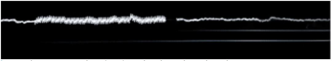
Figure 1: Change in processed EEG (pEEG) waveform during electrical stimulation.
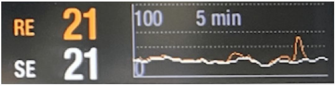
Figure 2: Change in spectral entropy waveform during electrical stimulation.
Discussion
EEG monitors brain function during anesthesia, providing invaluable real-time information about the patient’s neurophysiological status. This application of EEG is critical in ensuring patient safety and optimizing anesthetic delivery. By observing changes in EEG patterns, anesthesiologists can adjust the DoA, thereby minimizing awareness risks during surgery and the likelihood of postoperative cognitive dysfunction, especially in high-risk patient cohorts. EEG-directed anesthesia has demonstrated efficacy in reducing the amount of anesthetic required, expediting emergence from general anesthesia, and diminishing post-anesthesia care unit stays before discharge [3].
Gibbs et al. documented the impact of anesthetic agents on EEG in 1937, laying the groundwork for employing EEG in monitoring the DoA. From the 1990s onward, EEG has been extensively utilized in clinical settings to evaluate the DoA and sedation [4,5]. Since the inception of BIS monitoring in 1996, advancements in EEG monitoring techniques have been significant. EEG monitoring is instrumental in maintaining an optimal DoA, thereby reducing the anesthetic required. Each index was designed to simplify the interpretation of EEG data into a single numerical value that anesthesia clinicians could use clinically to make informed decisions about drug delivery and patient state.
Despite its benefits, EEG monitoring during anesthesia is often complicated by the presence of artifacts, which can significantly distort data. Artifacts during anesthesia can be broadly classified based on location. The most common cause of artifacts from the head but outside of the brain are Electromyographic (EMG) artifacts. EMG artifacts can arise from muscles outside the brain, such as the frontalis, masseter, or extraocular eye muscles. These artifacts can mimic high-frequency EEG activity and may be challenging to filter out completely. They can be distinguished by their frequency range (10-300 Hz) and characteristic patterns, such as frontalis «grimacing» [6].
The most common cause of artifacts from outside the head includes external interference. These artifacts are from electrical equipment, external pacemakers, ECG, diathermy, surgical movements, or power-line sources which can introduce noise into the EEG signal. For example, electrical noise from surgical equipment, such as cauterizing tools, can produce high-frequency signals that overlap with the gamma wave frequency band of EEG, often interpreted as a sign of inadequate anesthesia depth [7,8]. These artifacts pose a significant challenge as they can mimic or obscure the EEG signals, leading to misinterpretation of a patient’s neurophysiological state.
The pulsatile artifact is particularly challenging, which often occurs due to the rhythmic blood flow pressure that can affect the electrodes recording the EEG. For example, in a recent study 2021, Kamata et al. followed a 52-year-old patient undergoing general anesthesia for necrotizing fasciitis in the right femur with the DoA monitored with the SedLine® Brain Function Monitoring System [9]. The investigators found that EEG initially captured a rhythmic artifact synchronized with the heart rate, which was reduced after adjusting the electrodes. Further offline analysis indicated that this EEG interference matched the heart rate frequency. This case illustrates how a pulsation artifact from the superficial temporal artery can affect EEG signals during anesthesia monitoring.
In our cases, disturbances in perioperative EEG waveforms were observed during BAT, significantly changing algorithmically calculated RE and SE Entropy values. One reason for the EEG alterations could be cardiac impulses triggered by BAT, which are detected as a pulsating artifact in Entropy. Another likely explanation for these perturbations is the 40 Hz artifact related to electrical impulses used for carotid sinus stimulation impacting the EEG signal. Of importance, misinterpretation of the Entropy readings will likely also appear for subsequent anesthetics in similar patients, attributable to gradual adjustment in the amplitude requirements of the carotid sinus baroreflex activation device over several months, i.e., increasing mAmp currents at 40 Hz frequency. This progressive increase of the stimulating current upon the carotid sinus has been attributed to scarring around the electrodes, exposing the electrodes to further distortion of the EEG and preventing the use of this signal to gauge the DoA [10].
Conclusion
The adjustments to the baroreflex as part of the BAT therapy process can increase the left ventricular ejection fraction. The mechanisms for this still need to be understood. Vigilant monitoring and further research are crucial to better understanding BAT’s effects on perioperative EEG, possibly necessitating adjusted interpretation strategies.
References
- Yancy CW, et al. 2013 ACCF/AHA guideline for the management of heart failure: a report of the American College of Cardiology Foundation/American Heart Association Task Force on Practice Guidelines. J Am Coll Cardiol. 2013; 62: e147-239.
- Zile MR, et al. Baroreflex activation therapy for the treatment of heart failure with a reduced ejection fraction: safety and efficacy in patients with and without cardiac resynchronization therapy. Eur J Heart Fail. 2015; 17: 1066-74.
- Punjasawadwong Y, A Phongchiewboon, N Bunchungmongkol. Bispectral index for improving anaesthetic delivery and postoperative recovery. Cochrane Database Syst Rev. 2014; 2014: CD003843.
- Gibbs FA, El Gibbs, WG Lennox. Effect on the Electro Encephalogram ff Certain Drugs Which Influence Nervous Activity. Archives of Internal Medicine. 1937; 60: 154-166.
- Sun Y, et al. Electroencephalography: Clinical Applications During the Perioperative Period. Front Med (Lausanne). 2020; 7: 251.
- Bennett C, et al. Practical use of the raw electroencephalogram waveform during general anesthesia: the art and science. Anesth Analg. 2009; 109: 539-50.
- Cascella M. Mechanisms underlying brain monitoring during anesthesia: limitations, possible improvements, and perspectives. Korean J Anesthesiol. 2016; 69: 113-20.
- Guerit JM. Neuromonitoring in the operating room: why, when, and how to monitor?. Electroencephalogr Clin Neurophysiol. 1998; 106: 1-21.
- Kamata K, et al. Spurious electroencephalographic activity due to pulsation artifact in the depth of anesthesia monitor. JA Clin Rep. 2021; 7: 35.
- Zile MR, et al. Baroreflex Activation Therapy in Patients with Heart Failure with Reduced Ejection Fraction. J Am Coll Cardiol. 2020; 76: 1-13.
