
Open Access, Volume 10
Trichoscopic features in scalp dermatomyositis
M Lahouel; R Said El Mabrouk*; S Mokni; N Ghariani Fetoui; M Thabouti; S Saad; M Ben Rejeb; Belkahla; A Aounallah; N Ghariani; M Denguezli
Dermatology Department, Farhat Hached Hospital, University of Sousse, Tunisie.
Randa Said El Mabrouk
Dermatology Department, Farhat Hached Hospital, University of Sousse, Tunisie.
Tel: +216-54-49-22 41;
Email: saidranda@yahoo.fr
Received : May 15, 2024,
Accepted : June 07, 2024
Published : June 10, 2024,
Archived : www.jclinmedcasereports.com
Abstract
Scalp involvement in Dermatomyositis (DM) represents a diagnostic challenge. Its clinical presentation is often atypical, and can be observed in other collagenosis. In this report, we outline the trichoscopic signs of two cases of DM of the Scalp (SDM).
Trichoscopy revealed cicatricial alopecia (absence of follicular openings with white structureless areas), enlarged tortuous capillaries, linear branched vessels, curved branched vessels, lake-like vessels, perifollicular and interfollicular scales. We observed an uncommon sign that was the «large yellowish clods», corresponding to giant follicular plugs.
In the field of connective tissue diseases, dermoscopy gained popularity for the evaluation of inflammatory skin conditions. The clinico-dermoscopic correlation seems to be an accessible substitute for biopsy. Trichoscopy is a rapid, easy to apply, non-invasive technique that enables the diagnosis and should be performed routinely in all patients with dermatomyositis.
Keywords: Trichoscopie; Alopecia; Dermatomyositis.
Copy right Statement: Content published in the journal follows Creative Commons Attribution License (http://creativecommons.org/licenses/by/4.0). © Mabrouk RSE (2024)
Journal: Open Journal of Clinical and Medical Case Reports is an international, open access, peer reviewed Journal mainly focused exclusively on the medical and clinical case reports.
Citation: Lahouel M, Mabrouk RSE, Mokni S, Fetoui NG, Thabouti M, Saad S, Rejeb MB. Trichoscopic features in scalp dermatomyositis. Open J Clin Med Case Rep. 2024; 2253.
Introduction
Scalp involvement in Dermatomyositis (DM) represents a diagnostic challenge. Its clinical presentation is often atypical, and can be observed in other collagenosis, which can delay the diagnosis and consequently the treatment of this affliction. Its dermoscopic characteristics have been poorly described. In this report, we outline the trichoscopic signs of two cases of DM of the Scalp (SDM).
Case Presentation
A 56-year-old female patient was diagnosed with amyopathic dermatomyositis since 1998. She had a characteristic rash of dermatomyositis consisting in heliotropic periorbital erythema, Gottron’s papules, violaceous scaling patches in her upper extremities, and periungual erythema with telangiectasias. During the follow up, she never developed an inflammatory myopathy but had several cutaneous flare-ups. Repeated explorations did not show evidence of malignancy, infection, or association with another collagenosis. Topical corticosteroids, methotrexate, antimalarials lead to partial improvement. Recently, the patient developed persistent scalp itchiness. The scalp examination showed a large erythematous, squamous and telangiectasic alopecic plaque of the vertex (Figure 1). Trichoscopy revealed cicatricial alopecia (absence of follicular openings with white structureless areas), enlarged tortuous capillaries, linear branched vessels, curved branched vessels, lake-like vessels, perifollicular and interfollicular scales (Figure 2).
The second patient was a 65-year-old female patient who had been treated for DM since 2013. She presented with several erythematous and pruritic, alopecic plaques in the frontal, parietal and occipital areas. These lesions were associated with poikiloderma and had been evolving for 1 year. Trichoscopic examination revealed signs of scarring alopecia, blood vessels and perifollicular skin similar to the first patient, accompanied by a perifollicular hyperpigmentation.
In both our patients, we observed «large yellowish clods» (Figure 2), corresponding to giant follicular plugs. Mycological samples were negative, and scalp biopsies ruled out other diagnoses and confirmed the diagnosis of SDM.
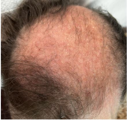
Figure 1: Alopecic plaque on the vertex.
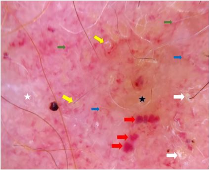
Figure 2: Linear branched vessels (green arrows), curved
branched vessels (bleu arrows), large yellow clods (yellow
arrows), white structureless area (white star), yellow structureless area (black star), lake-like vessels (red arrows).
Discussion/Conclusion
SDM mainly affects women. Clinically, it is characterized by alopecia, pruritus and/or a burning sensation [1]. These chronic scalp symptoms impair the patient’s quality of life and require adequate treatment. Trichoscopy is a non-invasive and helpful tool for the diagnosis of SDM. Jasso-Olivares and al. reported the biggest study describing clinical and trichoscopic features of 31 cases of SDM [1]. The most common findings included enlarged tortuous capillaries and peripilar scales. Tufting with three or more hair shafts emerging together, interfollicular scales, bushy capillaries, interfollicular and perifollicular pigmentation and vascular lake-like structures were least common. Zychowska et al. described other trichoscopic features of SDM such as yellow dots, hair diameter diversity, and interfollicular honeycomb pigment pattern [2]. The main differential diagnosis is discoid lupus with some dermoscopic similarities such as white dots and patches, brown honeycomb pattern, interfollicular scales, dotted vessels, arborizing vessels. However, enlarged capillaries with a tortuous shape, linear and curved branched vessels, and lake-like vessels seen in our patients seems to be more specific of SDM [3-5]. For our patients, trichoscopy showed specific findings of SDM. We also observed “large yellow clods” which were not previously described in the literature and they correspond to giant follicular plugs.
In the field of connective tissue diseases, dermoscopy gained popularity for the evaluation of inflammatory skin conditions [5]. The clinico-dermoscopic correlation seems to be an accessible substitute for biopsy. Trichoscopy is a rapid, easy to apply, non-invasive technique that enables the diagnosis and should be performed routinely in all patients with dermatomyositis.
References
- Jasso-Olivares, et al. Clinical and Dermoscopic Features of the Scalp in 31 Patients with Dermatomyositis, Skin Appendage Disord. 2017; 3: 119-124.
- Zychowska M, Reich A. Dermoscopy and Trichoscopy in Dermatomyositis-A Cross-Sectional Study. J. Clin. Med. 2022; 11: 375.
- Chanprapaph K, Limtong P, Ngamjanyaporn P, Suchonwanit P. Trichoscopic Signs in Dermatomyositis, Systemic Lupus Erythematosus, and Systemic Sclerosis: A Comparative Study of 150 Patients. Dermatology. 2021; 1-11.
- Chojer P, Mahajan BB. Nail fold dermo scopy in collagen vascular disorders: A cross-sectional study. Indian J. Dermatol. Venereol. Leprol. 2019; 85: 439.
- Joanna Golińska, Marta Sar-Pomian, Lidia Rudnicka. Diagnostic Accuracy of Trichoscopy in Inflammatory Scalp Diseases: A Systematic Review, Dermatology. 2022; 238: 412-421.
