
Open Access, Volume 10
Infrequent spontaneous fundus lesions in a Macaca fascicularis cohort from southern China: A case-series study
Lijie Pan1,2; Ningli Wang1,2; Wei Liu3,4; Zhiwei Huang3 ; Yehong Zhuo5 ; Jian Wu1,6*
1Henan Academy of Innovations in Medical Science, Zhengzhou, Henan 450000, China.
2Beijing Institute of Ophthalmology, Beijing Tongren Eye Center, Beijing Tongren Hospital, Capital Medical University, Beijing Ophthalmology & Visual Sciences Key Laboratory, Beijing 100730, China.
3Guangzhou Huazhen Biosciences, Guangzhou 510900, China.
4School of Food Sciences and Engineering, South China University of Technology, Guangzhou, 510641, China.
5State Key Laboratory of Ophthalmology, Zhongshan Ophthalmic Center, Sun Yat-sen University, Guangzhou 510060, China.
6School of Life Sciences, Peking University, Beijing 100871, China.
Jian Wu
Henan Academy of Innovations in Medical Science, Biotechnology Street, Hang kong Gang District, Zhengzhou, Henan, China.
Email: karena.wu@foxmail.com
Received : May 05, 2024,
Accepted : May 28, 2024
Published : May 31, 2024,
Archived : www.jclinmedcasereports.com
Abstract
Introduction: Non-Human Primates (NHPs) are suitable for studying human visual biology and ocular diseases since they share almost identical anatomic structure and biological function. From this eye study of a NHP cohort, some infrequent fundus lesions were shown and ideal animal models were provided for further exploration on pathogenesis and potential treatments of these diseases.
Methods: A cross-sectional ocular examination study of Macaques with the age ranged from 2 to 26 years was held in Guangdong Province (southeastern China) from 2021 to 2022. We acquired systematic data and ocular data from young, middle-aged to old individuals, including measurement of Intraocular Pressure (IOP), slit-lamp examination, Color Fundus Photograph (CFP), spectral domain- Optical Coherence Tomography (OCT), etc. Abnormal manifestations in fundus photograph, macular and optic OCT imaging were chosen for further discussion, and the retinal parameters beyond normal reference were also highlighted.
Results: Of all the individuals qualified for our research, some infrequent fundus diseases rather than those more common including high myopia, Age-Related Macular Degeneration (AMD) and glaucoma were observed, such as Myelinated Retinal Nerve Fibers (MRNFs), various types of chorioretinal atrophy, fundus flavimaculatus, optic atrophy, proliferative vitreoretinopathy, retinal vascular disease and so on. The fundus characteristics presented shared highly similarities from that of human.
Conclusion: NHPs are ideal animal models for preclinical research of some fundus diseases, thus contributing to further exploration on pathogenesis and potential treatments of fundus lesions.
Keywords: Non-human primates; Fundus lesion; Myelinated retinal nerve fibers; Chorioretinal atrophy.
Abbreviations: NHP: Non-Human Primate; IOP: Intraocular Pressure; CFP: Color Fundus Photograph; OCT: Optical Coherence Tomography; AMD: Age-Related Macular Degeneration; MRNF: Myelinated Retinal Nerve Fibers.
Copy right Statement: Content published in the journal follows Creative Commons Attribution License (http://creativecommons.org/licenses/by/4.0). © Wu J (2024)
Journal: Open Journal of Clinical and Medical Case Reports is an international, open access, peer reviewed Journal mainly focused exclusively on the medical and clinical case reports.
Citation: Pan L, Wang N, Liu W, Huang Z, Zhuo Y, Wu J. Infrequent spontaneous fundus lesions in a Macaca fascicularis cohort from southern China: A case-series study. Open J Clin Med Case Rep. 2024; 2248.
Introduction
A majority of fundus diseases are the contribution causes of visual impairment worldwide [1]. It was estimated that there were 43.3 million blind individuals worldwide in the year 2020, of which glaucoma optic neuropathy, age-related macular degeneration and diabetic retinopathy were all among the leading causes [2]. What’s more, with the development of health management and elongation of life expectancy, the occurrence of fundus diseases would see a gradually increase, putting substantial stress in the life quality for elder groups and the economic burden for the society as well.
Animal models have promoted ophthalmological research for decades of years as a result of their simulation to physiological condition in vivo [3]. Among them, Non-Human Primates (NHPs) are undoubtedly suitable and precise models for human visual biology and ocular diseases since they share almost identical anatomic structure and biological function, making it difficult to distinguish fundus images of NHPs from human even for experienced ophthalmologists.
Up to now, numerous studies have been launched about ocular parameters and features of some common diseases in NHP cohorts. However, other infrequent fundus disorders should not be neglected at all. From our NHP eye study held in Guangdong Province from 2021 to 2022, 395 subjects were incorporated into data analysis, and except for those spontaneous ocular diseases with high incidence, some lesions were hard to be defined or clarified explicitly, which appealed to further investigation and discussion.
Case Presentation
Myelinated Retinal Nerve Fibers (MRNFs)
Case 1: The 6.65-year-old male monkey was diagnosed with myelinated retinal nerve fibers in his right eye with typical fundus findings. The white myelin patch was grossy and opaque with feather-like margins, distributed around the optic disc and extended along the arrangement of nerve fibers (Figure 1A-B). No other obvious abnormality in the retina could be noticed. The dioptric condition as well as axial length for both eyes were within the normal range.
The other two cases were shown in Supplementary Figure 1.
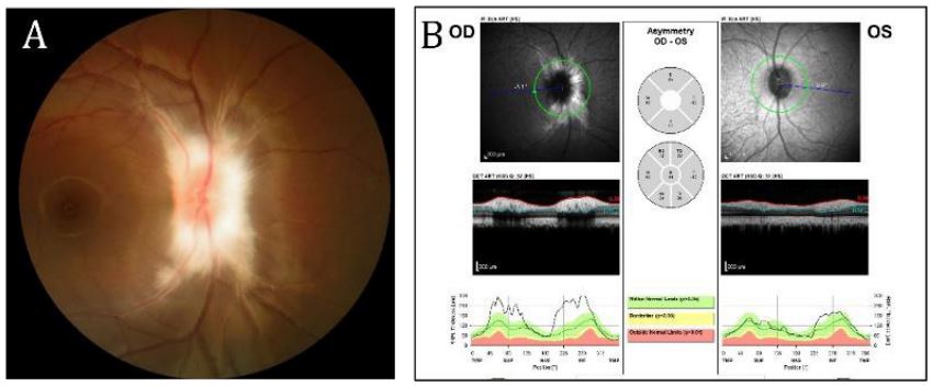
Figure 1: (A) Color Fundus Photography (CFP) of the right eye, showing the white
myelin patch in the juxtapapillary location; (B) Correlating Optical Coherence Tomography (OCT) showed a highly reflective myelinated inner retinal layer.
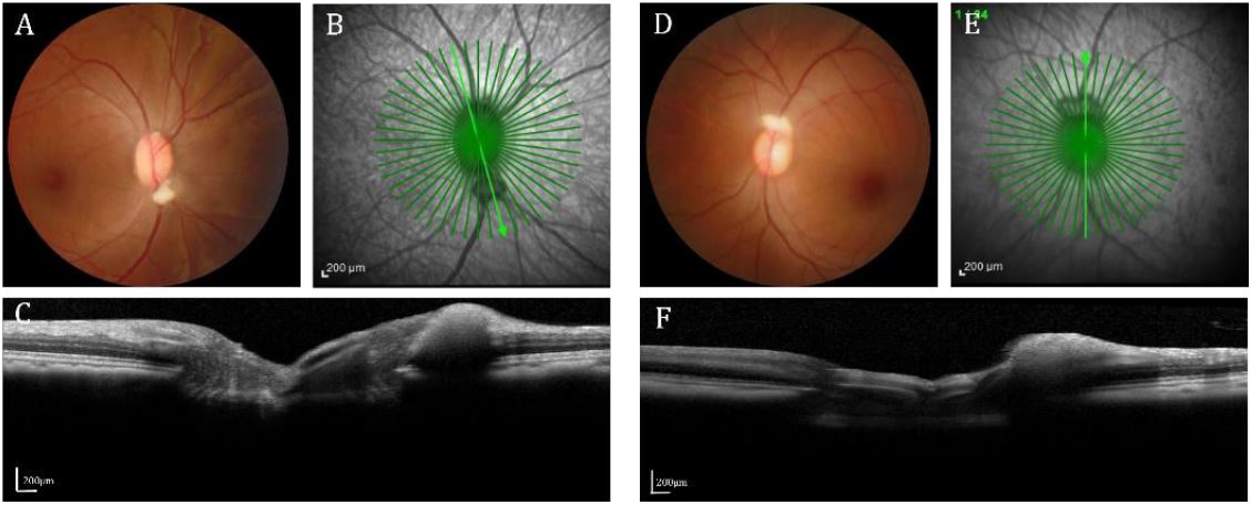
Supplementary Figure 1: (A) CFP of the right eye of the case showing the yellow-white lesions separately on the superior, temporal and inferior margin of the optic disc; (B-C) OCT
showed focal elevation of retinal nerve fiber layer near the disc. (D) CFP of the other case
showing the lesion in the superior of the optic disc in the left eye; (E-F) OCT showed focal elevation of retinal nerve fiber layer near the disc. CFP: Color Fundus Photography, OCT: Optical
Coherence Tomography.
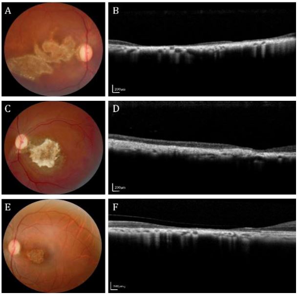
Figure 2: Appearance of various types of chorioretinal atrophy in the cynomolgus monkeys.
(A, C and E) CFPs showing irregular atrophy patches in local retina; (B, D and F) Linear OCT
scan of the lesions showing attenuation of RNFL, RPE/Bruch’s membrane complex and choroid
with impaired vague fundus structure. CFP: Color Fundus Photography, OCT: Optical Coherence Tomography; RNFL: Retinal Nerve Fiber Layer.
Chorioretinal atrophy
Cases 2-4: Case 2, 3 and 4 were separately from three 18-year-old monkeys (Figure 2A-F). The irregular atrophy patches could be seen mainly in the inferotemporal region of the retina, with relatively clear boundaries and irregular pigmentation deposition. Optical Coherence Tomography (OCT) showed attenuation and atrophy of both inner and outer retinal layers as well as the choroid. Since the medical history of the animals were hard to trace back, we speculated possible pathogenesis of chronic inflammation for case 2, 4 and trauma or Age-Related Macular Degeneration (AMD) for case 3.
Fundus flavimaculatus
Case 5: It’s a 12-year-old female monkey whose binocular fundus were full of irregular yellow-white spots especially in the inferior and temporal retina, some were oval, round, linear while others were fishtail- or tadpole-shaped (Figures 3A and B). Atrophy arc and pigment disorder were seen around the left optic disc (Figure 3B). OCT image of the left eye showed focal hyperreflection and deformation in the internal layers of retina, which may be resulted from excessive lipofuscin deposition (Figure 3C).
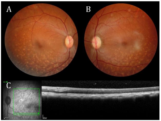
Figure 3: (A and B) Binocular CFPs showing a large amount of irregular yellow-white flecks
almost around the whole retina, especially in the inferior and temporal parts. Central fovea
were not affected yet. (C) Linear OCT scan of the lesions showing a slightly deformation and
hyperreflection of the inner retina. There were no obvious pathological alterations in the RPE
layer. CFP: Color Fundus Photography, OCT: Optical Coherence Tomography
We also identified other fundus lesions such as optic nerve atrophy with unknown pathogenesis and proliferative vitreoretinopathy (PVR), whose color fundus photography and OCT images were provided in Supplementary (Figures 2 and 3).
Discussion
In this study, we carried out the comprehensive examinations for 395 Macaca fascicularis. Of all the individuals qualified for our research, some infrequent fundus diseases were observed such as MRNFs (3 cases), various types of chorioretinal atrophy (7 cases), fundus flavimaculatus (1 case), optic atrophy (2 cases), proliferative vitreoretinopathy (1 case), retinal vascular diseases (3 cases, not shown) and so on.
MRNFs are a congenital benign lesion characterized by myelination of retinal ganglion cell axons passing through the lamina cribrosa of optic nerve head, and thus appearing as a white thickening patch with feathered borders in the inner layer of retina, obscuring the underlying tissue. The incidence of MRNFs in our study is 0.76%, consistent with the reported 0.4%-1.0% in general population [4], which reveals the correspondence in the epidemiology of fundus lesions between human and cynomolgus and lays a foundation for taking advantage of these animal models for further research.
Of all the cynomolgus investigated, chorioretinal atrophy was observed in 7 cases without obvious causes such as AMD, pathologic myopia and chorioretinitis, for a prevalence of 1.77%. It’s hard for us to compare the incidence with other primate studies since it seems to be sporadic and multi-factor induced. Monkeys in our study are raised by artificial facilities in relatively small rooms supplied with appropriate temperature, humidity and nutrients, which are different from seminatural free-ranging surroundings in light exposure intensity, accident rate as well as immunity condition.
Fundus flavimaculatus shares some similarities with Stargardt Disease (STGD1) which would cause progressive macular dystrophy in juvenile due to mutations in ABCA4 [5]. But macula is typically not involved in fundus flavimaculatus. The case presented in our study could correspond to it for multiple regular white-yellow flecks can be found scattered in periphery retina. Unfortunately, Fluorescein Fundus Angiography (FFA) and genetic detection have not been performed during the follow-up of the monkey. It would be interesting to complete the study in search of more evidence to confirm the diagnosis in the future.
There are some limitations of this study that should be discussed. First, this was a relatively small case series of Macaca fascicularis seen in our NHP eye study conducted in southern China and may not reflect the disease spectrum of other NHP cohorts. Furthermore, some important examinations which were likely to help for clinical diagnosis were missed due to some unpredicted reasons like unavailability of examination devices, animal safety and cost-effectiveness and so on.
In conclusion, we conducted the fundus screening in a large NHP cohort in Guangzhou, China and summarized some infrequent spontaneous lesions in cynomolgus from different ages resembling human diseases, thus providing ideal animal models with partly specific and known environmental exposure and pedigree for preclinical research for further exploration on pathogenesis and potential treatments.
Declarations
Ethics approval and consent to participate: This study adhered to the National Institutes of Health Guide for the Care and the guidelines of the Association for Research of Vision and Ophthalmology for the Use of Animal in Ophthalmic and Vision Research. This study was approved by the Ethical Committee of the Guangzhou Huazhen Biosciences Company (Ethics Number: 2020-168) and Zhongshan Ophthalmic Center (Permit Number: SYXK (YUE) 2018-0189).
Authors’ contributions: Lijie Pan, Ningli Wang and Jian Wu completed the study design and data collection. Lijie Pan completed image processing and manuscript writing. Wei Liu, Zhiwei Huang and Yehong Zhuo participated in the data collection. Jian Wu was responsible for revising the draft.
Financial support: This study was supported by grants from the National Key R&D Project of China (2020YFA0112701), the National Natural Science Foundation of China (82171057), Science and Technology Program of Guangzhou, China (202206080005), and the National Natural Science Foundation of China (GZR-2012–009).
Conflict of interest: No conflicting relationship exists for any author
References
- Blindness GBD, Vision Impairment C. Vision Loss Expert Group of the Global Burden of Disease S. Causes of blindness and vision impairment in 2020 and trends over 30 years, and prevalence of avoidable blindness in relation to VISION 2020: the Right to Sight: an analysis for the Global Burden of Disease Study. Lancet Glob Health 2021; 9(2): e144-e160. doi: 10.1016/S2214-109X(20)30489-7.
- Blindness GBD, Vision Impairment C. Vision Loss Expert Group of the Global Burden of Disease S. Trends in prevalence of blindness and distance and near vision impairment over 30 years: an analysis for the Global Burden of Disease Study. Lancet Glob Health 2021; 9(2): e130-e143. doi: 10.1016/S2214-109X(20)30425-3.
- Lin KH, Tran T, Kim S, Park S, Stout JT, et al. Advanced Retinal Imaging and Ocular Parameters of the Rhesus Macaque Eye. Transl Vis Sci Technol 2021; 10(6): 7. doi: 10.1167/tvst.10.6.7.
- Teixeira F, Fonseca AC, Pinto F. Acquired and progressive myelinated retinal nerve fibers in neurofibromatosis type 1. J AAPOS. 2019; 23 (3): 178-179. doi: 10.1016/j.jaapos.2019.01.012.
- Fujinami K, Zernant J, Chana RK, Wright GA, Tsunoda K, et al. Clinical and molecular characteristics of childhood-onset Stargardt disease. Ophthalmology. 2015; 122(2): 326-334. doi: 10.1016/j.ophtha.2014.08.012.
