
Open Access, Volume 10
Metabolic superscan on F-18 FDG PET/CT
Utkun Moran1; Doğangün Yüksel1*; Burcu Yapar Taşköylü2
1Department of Nuclear Medicine, Pamukkale University, Medical Faculty, Denizli, Türkiye.
2Department of Medical Oncology, Pamukkale University, Medical Faculty, Denizli, Türkiye.
Doğangün Yüksel
Department of Nuclear Medicine, Pamukkale University, Medical Faculty, Denizli, Türkiye.
Email: dyuksel@pau.edu.tr
Received : Jan 06, 2024,
Accepted : Mar 01, 2024
Published : Mar 11, 2024,
Archived : www.jclinmedcasereports.com
Abstract
Superscan is a classic finding identified especially in bone scintigraphy. It is generally seen in metabolic bone diseases and diffuse bone metastases. This case is presented to emphasize the distinction of malignbenign of the superscan in F-18 FDG PET/CT imaging.
Keywords: Pet; Fdg; Superscan; Osteodystrophy.
Abbreviations: FDG: Fluorodeoxyglucose; MIP: Maximum Intensity Projection; FDG PET/CT: Fluorodeoxyglucose Positron Emission Tomography/Computed Tomography.
Copy right Statement: Content published in the journal follows Creative Commons Attribution License (http://creativecommons.org/licenses/by/4.0). © Yüksel D (2024)
Journal: Open Journal of Clinical and Medical Case Reports is an international, open access, peer reviewed Journal mainly focused exclusively on the medical and clinical case reports.
Citation: Moran U, Yüksel D, Taşköylü BY. Metabolic super scan on F-18 FDG PET/CT. Open J Clin Med Case Rep. 2024; 2205.
Introduction
Superscan is a classic finding identified especially in bone scintigraphy. It is generally seen in metabolic bone diseases and diffuse bone metastases. This finding is characterized by diffuse homogeneous bone uptake of biphosphonate radiopharmaceuticals and lack of activity uptake in the kidneys in metabolic diseases. Metabolic bone diseases in which super scanning is defined are Paget’s disease, hyperparathyroidism, rickets, osteopetrosis, osteoporosis [1]. Diffuse bone metastases are characterized by heterogeneous skeletal system involvement and lack of activity involvement in the kidneys. It is usually seen in prostate cancer and breast cancer [1].
On the other hand, a similar finding in F-18 Fluorodeoxyglucose (FDG) Positron Emission Tomography/ Computed Tomography (F-18 FDG PET/CT) imaging has been described in very few publications [2-5]. It has been described in cancer cases on F-18 FDG PET imaging in the accessible literature. The superscan finding defined by F-18 FDG PET is similar to bone scintigraphy [2-5].
We share the superscan from metabolic bone disease/renal osteodystrophy of a female patient with breast cancer and chronic renal failure observed on F-18 FDG PET/CT scan.
Case Report
A 39 years old female patient with breast cancer was sent for the evaluation of post chemotherapy. The patient, who receives regular chemotherapy treatment (paclitaxel), has been following up with routine F-18 FDG PET/CT controls. Our patient has been undergoing hemodialysis treatment 3 times a week since 2012 due to idiopathic chronic kidney disease. Tc-99m MIBI dual phase parathyroid scintigraphy was performed on the patient due to previously elevated PTH on 2019. Parathyroid hyperplasia was detected in parathyroid scintigraphy (Figure 1). Before the F-18 FDG PET/CT scan, calcium, phosphorus, PTH and Dual Energy X-Ray Absorption meter (DEXA) measurements were performed on the patient by the oncology department. While calcium measurement values (last value: 11.26 mg/dl; normal range: 8.6-10.2 mg/dl) were generally high level, phosphorus measurement values (last value: 4.2 mg/dl; normal range: 2.6-4.5 mg/dl) were close to the upper limit of normal. The last PTH value was above 5,000 ng/L (normal range: 15-65 ng/L). The result of DEXA was compatible with osteopenia (t score vertebrae:-1.5 SD, femur:-1.7 SD).
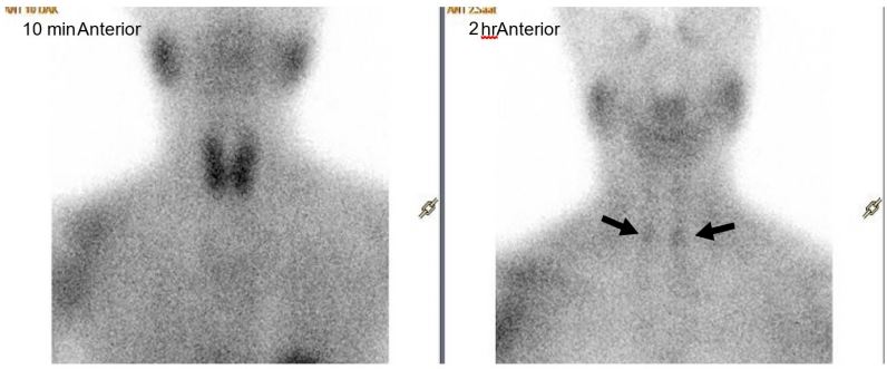
Figure 1: Dual-phase 99m Tc MIBI scintigraphy imaging with 740 MBq (20 mCi). The scintigraphy was taken with an anteriorposterior parallel hole collimator. Images were acquired at 10 minutes and 2 hours. That activity uptake was observed in the
lower lobes of the thyroid gland in the late images in the areas indicated by the arrow. Interpreted in favor of parathyroid
hyperplasia.
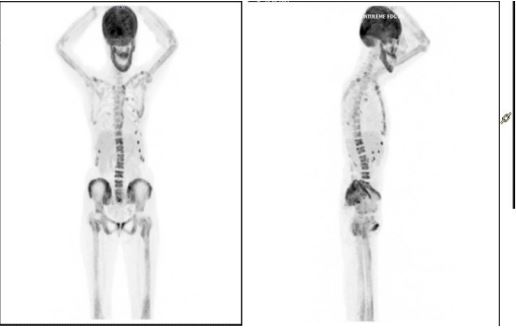
Figure 2: Whole body anterior (a) and lateral (b) Maximum Intensity Projection (MIP) images from F-18 Fluorodeoxyglucose Positron Emission Tomography/Computed Tomography (FDG PET/CT) scan show diffusely increased FDG uptake in
the costocondral joints without any abnormalities in bone structure, and absence of radiotracer excretion in the kidneys and
bladder, like the superscan finding to be described for a bone scan.
During the examinations of the patient who was added to the list for renal transplantation, a nodul was found in her right breast. Ductal carcinoma was found as a result of the biopsy pathology.
The patient underwent the modified radical mastectomy and lymphadenectomy after diagnosis. He then received chemotherapy and radiotherapy. She received radiotherapy 3 months and paclitaxel chemotherapy 8 months before the F-18 PET-CT imaging. We observed a super scan sign in the F-18 FDG PET/CT imaging. F-18 FDG showed homogeneous uptake in the skeletal system and the kidneys could not be clearly observed (Figure 2). Therefore, we thought that the super scan finding was due to metabolic bone disease rather than breast cancer metastasis.
The superscan sign which is detected by the F-18 FDG PET/CT imaging image is rarely observed. Its most common causes are in myeloproliferative diseases, metastatic diseases and metabolic bone diseases [5,6]. In the metastatic superscan due to extensive bone metastasis, while it is characterized by widespread irregular F-18 FDG uptake in the skeletal system, the bladder and kidneys were not observed to the accumulation of F-18 FDG PET/CT [1]. However, PET/CT superscan due to metabolic bone disease is characterized by widespread, homogeneous increased FDG uptake, as in bone scintigraphy (Figure 2).
When we looked laboratory results, it was seen that parathyroid hormone levels were always high. Hyperplasia on parathyroid scintigraphy and diffuse bone involvement on F-18 FDG PET/CT imaging support this diagnosis. It differed from the metastatic superscan appearance with an increase in diffuse homogeneous skeletal system uptake.
This case is presented to emphasize the distinction of malign-benign of the superscan in F-18 FDG PET/CT imaging.
References
- Fred A. Mettler, Milton J. Guiberteau. Essentials of Nuclear Medicine and Molecular Imaging. 7th edn. 244.
- Dae-Weung Kim, Chang Guhn Kim, Soon-Ah Park, Sang-Ah Jung, Sei-Hoon Yang. Metabolic super scan in 18F-FDG PET/CT imaging. Journal of Korean medical science. 2010. https://doi.org/10.3346/jkms.2010.25.8.1256
- Güney Isa Burak, Paydas Semra, Balli Huseyin Tugsan. Super Scan Caused by Parathyroid Carcinoma Observed Both in 18FFDG PET/CT Scan and Tc-99m MDP Bone Scintigraphy. Molecular Imaging and Radionuclide Therapy. 2017; 26(3). https://doi. org/10.4274/mirt.70188
- N Ghesani, Jin Jung, Shyam Patel, Tekchand Ramchand. Superscan caused by renal osteodystrophy: Observed on 18F FDG PET/ CT scan. Indian Journal of Nuclear Medicine. 2013. https://doi.org/10.4103/0972-3919.121981
- Ismaheel Lawal, Alfred Ankrah, Kehinde Ololade, Moshe Modiselle, Mike Sathekge. MD Renal osteodystrophy presenting as a metabolic superscan on F-18 FDG PET/CT. Medicine (Baltimore). 2017; 96(46): e8471. https://doi.org/10.1097/ MD.0000000000008471
