
Open Access, Volume 10
Obturator dislocation of the hip
Zied Mansi1*; Abdelkader Tounsi1; Aymen Ben Mahmoud1; Ben Jaballah Alaa1; Wajdi Chermiti2; Haggui Ali3; Zaidi Bacem4
1Department of Orthopedic Surgery, Ibn El Jazzar University Hospital, Kairouan, Tunisia.
2Department of Orthopedic Surgery, Sahloul University Hospital, Sousse, Tunisia.
3Department of Orthopedic Surgery, Hospital of Kasserine, Kasserine, Tunisia.
4Department of General Surgery, Ibn El Jazzar University Hospital, Kairouan, Tunisia.
Zied Mansi
Department of Orthopaedic Surgery, Ibn El Jazzar Kairouan University Hospital 3100 Kairouan, Tunisia.
Email: doc.zm@htmail.fr
Received : Jan 24, 2024,
Accepted : Feb 26, 2024
Published : Feb 28, 2024,
Archived : www.jclinmedcasereports.com
Abstract
Obturator dislocation of the hip is caused by high-velocity accidents as evidenced by its frequent association with other traumatic injuries and, seldom found. Its main complication remains femoral head avascular necrosis. We report on four cases of obturator dislocation of the hip. The mean age of patients was 30 years, and all their injuries followed a road traffic accident. Associated lesions were a contralateral femur fracture in two cases and an osteochondral fracture in one case. Reduction of dislocations was achieved orthopedically under general anaesthesia and the average waiting time before reduction was 20 hours. One patient had an intra-articular incarcerated frag- ment visible on X-ray, and another patient showed signs of early coxarthrosis 15 months later. The average follow-up time was 24 months.
Keywords: Dislocation; Anterior; Obturator; Hip; Adult.
Copy right Statement: Content published in the journal follows Creative Commons Attribution License (http://creativecommons.org/licenses/by/4.0). © Zied M (2024)
Journal: Open Journal of Clinical and Medical Case Reports is an international, open access, peer reviewed Journal mainly focused exclusively on the medical and clinical case reports.
Citation: Mansi Z, Tounsi A, Mahmoud AB, Alaa BJ, Chermiti W, Ali H, Bacem Z. Obturator dislocation of the hip. Open J Clin Med Case Rep. 2024; 2204.
Introduction
Table 1: Comparative table of the four dislocations.
| Patients | Age | Sex | Etiology | Dislocation type |
Associated lesions |
Reduction time (hours) |
Complications | Follow up (months) |
|---|---|---|---|---|---|---|---|---|
| 1 | 29 | M | RTA | Obturator |
Contralateral left
upracondylar open fracture |
18 | None | 39 |
| 2 | 26 | M | RTA' | Obturator | None | 24 | None | 24 |
| 3 | 33 | M | RTA' | Obturator | Left subtrochanteric fracture | 12 |
Coxarthrosis and
peri-articular ossifications |
15 |
| 4 | 33 | M | RTA | Obturator | None | 26 | None | 20 |
*Road traffic accident.
Traumatic anterior dislocation of the hip is an uncommon injury compared with posterior disloca- Traumatic anterior dislocation of the hip is an uncommon injury compared with posterior dislocation. It accounts for 5%-18% of all hip dislocations and, obturator dislocation is rare [1,2]. These injuries may occur in accidents by deceleration, in that the vehicle occupant is with his legs bent, abducted, and externally rotated during impact, as well as in motorcycle accidents in which the legs are often in hyper abduction. Anterior dislocations are subdivided into the superior type (pubic) and the inferior type (obturator). Non-operative closed reduction is the treatment of choice for this injury [1-5]. Obturator dislocations are serious injuries, associated with major lesions and occur in poly-traumatic contexts [6]. Their prognosis is mainly functional and linked to femoral head avascular necrosis and coxarthrosis [7-10] we report on 4 cases of obturator dislocation of the hip with an emphasis on the treatment and the prognosis.
Case 1: Mr. BA was a 29-year-old patient who was hospitalized in October 2000 following a road traffic accident. He was sitting behind the driver when his right knee hit the driver’s seat. On admission, the same limb was abducted, externally rotated and flexed at the hip. An anteroposterior radiograph of the pelvis revealed a right obturator dislocation (Figure 1) associated with a left supracondylar open fracture. Treatment consisted of closed reduction. The hip was stable afterward. The left supracondylar open fracture was treated surgically. Weight bearing on the dislocated limb was authorized after 6 weeks of bed traction. Thirty nine months later, the right hip is mobile, painless and normal on plain X-rays.
Case 2: Mr. CA was a 26-year-old patient who was hospitalized on February 7th 2004 for a right hip injury following a road traffic accident. He had no medical history and in the circumstances of the accident, he was carrying his cart from the front when a car hit the back of his cart. Immediately, he felt a sharp pain and an absolute functional impotence of the limb. Clinical examination of the same limb revealed abduction, an external rotation and flexion at the hip. Standard radiographs confirmed obturator dislocation (Figure 2). Closed reduction under general anaesthesia was achieved 24 hours after the injury, followed by 10 days of bed traction. Control plain X-rays obtained 24 months later showed no signs of avascular necrosis.
Case 3: SA was 33 with no medical history, when he was referred to us the 17/09/2005; for the management of multiple injuries following a road traffic accident. On September 16th 2005, the patient was riding a motorbike when he fell off trying to avoid potholes. Initial clinical findings on arrival noted: swelling and sharp pain on the superior one-third of the thigh to the left associated with a rotated foot and a skewed patella; abduction, external rotation and flexion at the hip to the right lower limb.
Plain X-rays confirmed a left subtrochanteric fracture and a right obturator dislocation of the hip with an intra-articular incarcerated fragment (Figure 3). Closed reduction of the dislocated hip and surgical treatment of the left subtrochanteric fracture by a Küntscher intramedullary nail; were performed the same day the patient was admitted. Post-reduction radiographs showed an articular gap widening with a defect in the superior lateral femoral head (Figure 4). A confirmation CT scan demonstrated an intra-articular incarcerated fragment (Figure 5). An arthroscopic extraction was warranted, but due to inadequate hospital equipment and patient financial hardship; the patient did not receive appropriate care. Fifteen months later; Mr. SA presented with a control X-ray showing early signs of coxarthrosis and peri-articular ossifications (Figure 6).
Case 4: Mr. DS was 31 with no medical history, when he was referred to us the 06/09/2013; for a left hip injury follow- ing a road traffic accident. On September 6th 2013, the patient was riding a motorbike when he collided with another motorbike rider, hitting the left knee. On admission, clinical examination revealed abduction, external rotation and flexion at the hip to the same limb. Plain X-rays showed obturator dislocation of the hip (Figure 7). Treatment consisted of closed reduction the next day, followed by 15 days of bed traction. A control MRI 20 months post-traumatic showed no signs of avascular necrosis of the femoral head (Figure 8).
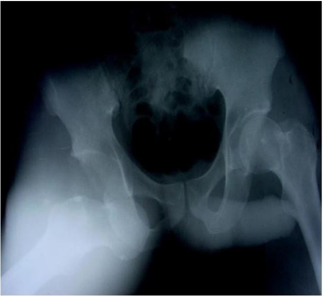
Figure 1: Anteroposterior X-ray of the pelvis showing a
right obturator dislocation (patient 1).
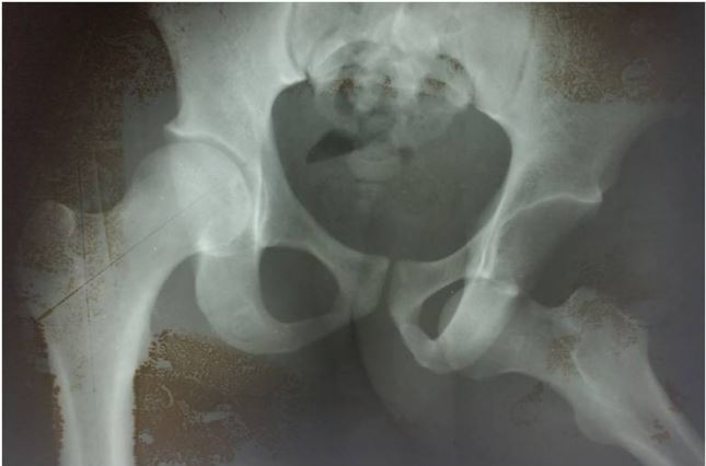
Figure 2: Anteroposterior X-ray of the pelvis showing a
left obturator dislocation (patient 2).
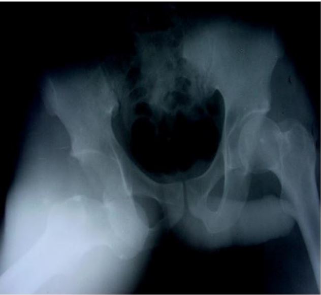
Figure 3: Anteroposterior X-ray of the pelvis showing a
right obturator dislocation with an intra-articular incarcerated fragment (arrow) and a left subtrochanteric fracture (patient 3).
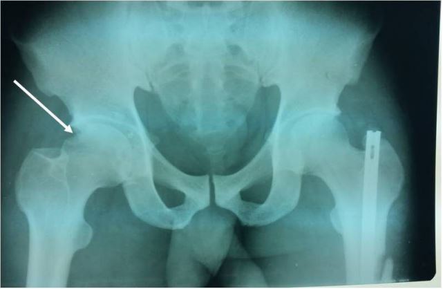
Figure 4: Anteroposterior X-ray of the pelvis showing an
articular gap widening with a defect in the superior lateral femoral head (patient 3).
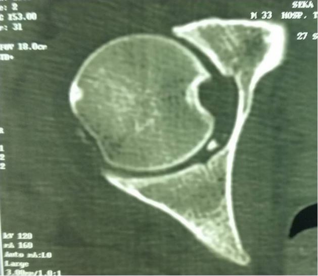
Figure 5: CT scan showing an intra-articular fragment
(patient 3).
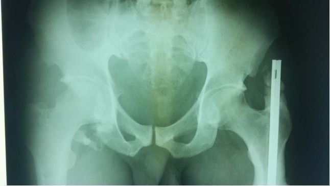
Figure 6: Anteroposterior X-ray showing coxarthrosis
and peri-articular ossifications (patient3).
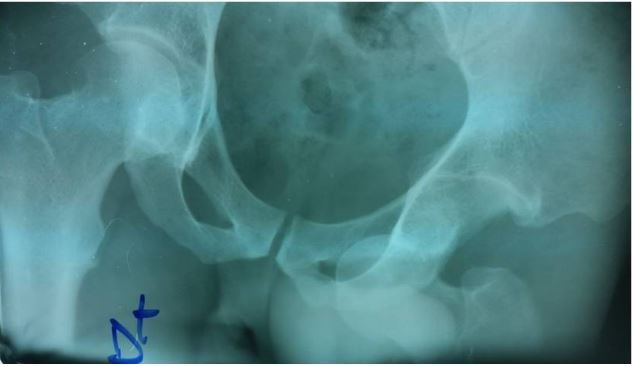
Figure 7: Anteroposterior X-ray of the pelvis showing a
left obturator dislocation (patient 4).
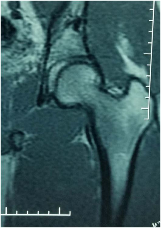
Figure 8: MRI showing no signs of avascular necrosis of
the femoral head (patient 4).
Discussion
The hip joint is inherently stable, which requires significant force to promote its dislocation. Thus, hip dislocations usually result from high-energy trauma. The position of the femoral head in relation to the acetabulum and the vector of the force determine the type of injury produced. Hence, the high predominance of posterior dislocations compared with anterior ones; as the postero-inferior capsule constitutes the weakest link [3-5].
Anterior dislocations of the hip account for 5%-18% of all hip dislocations and require high-velocity accidents with an impact area located at the internal surface of the flexed knee [3-5]. The angle of hip flexion will explain the type of anterior dislocation; superior (pubic) or inferior (obturator). There is strong evidence to support that simultaneous abduction, external rotation and flexion of the hip are necessary before an obturator dislocation can occur [2,4,5,7]. In two of our cases, the presence of multiple lesions is consistent with high- velocity trauma [3,4,6,10].
The striking deformity of the limb makes the diagnosis easy in Accident & Emergency units. Confirmation is made using standard radiographs. Clinical examination must look for vascular complications; since cases of anterior dislocations associated with these lesions have been reported in the literature [6,10,11]. Anterior capsular lesions (tearing or disinsertion) are permanent features. There is a frequent association with osteoarticular lesions, osteochondral femoral head fractures being the most common, as seen in one of our patients. A CT scan must be warranted to confirm those lesions, reveal intra-articular incarcerated fragments and better appreciate any association with a pelvic brim fracture as in one of our patients [12-14].
Obturator dislocation, as all hip dislocations, is an orthopedic emergency which requires immediate attention, so to enhance the functional prognosis. However, our average waiting time before reduction of 20 hours, reduction must be achieved ideally within 6 hours post-traumatic. Delays recorded in our cases could be explained by the design of our health care system, in which patients foot the bill for their care. Closed reduction is achieved under general an aesthesia, in theatre, as in all our patients; using zenith traction, adduction and internal rotation technique. Surgical reduction is warranted if closed reduction fails. Hip stability testing following reduction is paramount; so is the success of reduction which needs to be appreciated by control fluoroscopy and standard radiographs [2,4,8].
Cranio-cerebral, thoracic, and abdominal injuries are commonly associated. Skeletal injuries often associated include fractures of head or femoral neck, femoral shaft, acetabulum and pelvis, as well as knee, ankle and foot injuries and neurological lesion. Apart from life threatening injuries, dislocation reduction should take precedence over treatment of all other skeletal injuries [3,6].
Asymmetric articular gap widening of the injured hip joint is pathognomonic of capsular interposition or intra-articular incarcerated fragment, until proven otherwise. Arthroscopic extraction must be warranted [15,16]. Failure to treat this complication could prove catastrophic, as the unexpected appearance of early coxarthrosis in one of our patients.
There is no consensus on the length of bed traction and of non-weight bearing after reduction. Some authors advocate for a few days of pain management traction and 3-6 weeks of non-weight bearing [2,3,11,17]. A contralateral femur fracture prolonged non-weight bearing after three weeks of bed traction in 2 of our patients, as supported by Dawson and Van Rijn [1]. The attitude in these cases will depend on associated lesions [12,17].
Clinical and radiological evidence of avascular necrosis will take at least 17 months before being noticed [9,18]. A minimum follow-up of 2 years is required before reviewing the long term outcome; such as femoral head avascular necrosis, peri-articular ossifications and coxarthrosis [1,4]. Avascular necrosis is directly linked to the timing of dislocation reduction and treatment of associated lesions, which should be within 6 hours post-traumatic [3,14]. However, our average waiting time before reduction of 20 hours, we recorded only one case of coxarthrosis following untreated intra-articular incarcerated fragment. Coxarthrosis is the major dreadful long-term risk; and its incidence is difficult to evaluate, due to the lack of follow-up and small size of studies reported in the literature. Coxarthrosis intimately follows necrosis. Hougaard and Thomsen [9] reported a 4.8% incidence of avascular necrosis of the femoral head when the hip was reduced within 6 hours, compared to an incidence of 58.8% when reduced after 6 hours. Generally speaking, outcome deteriorates as time elapses, re- sulting up to 90% of arthrosis [9].
The follow-up of femoral head necrosis consists of bone scintigraphy or better MRI of the hip [19,20].
Conclusion
Obturator dislocation of the hip is an orthopedic emergency, easy to recognize clinically and radiologically in Accident & Emergency units; however, it is seldom found. The mechanism of injury could explain its frequent association with other traumatic lesions. Treatment consists most of the time of closed reduction under general anesthesia. Femoral head avascular necrosis remains the main complication, and its follow-up requires repeated bone scintigraphy or better MRI.
Conflict of interest: None.
References
- Dawson I, Van Rijn ABB. Traumatic Anterior Dislocation of the Hip. Archives of Orthopedic and Trauma Surgery. 1989; 108: 55-57. http://dx.doi.org/10.1007/BF00934160
- Shimi M, El Idrissi M, Dahmani O, et al. La luxation antérieure obturatrice pure de la hanche. À propos de 2 case. Tunisie Orthopédique. 2010; 3: 73-75.
- Clegg TE, Roberts CS, Greene JW, et al. Hip Dislocations-Epidemiology, Treatment, and Outcomes. In- jury. 2010; 41: 329-334. http://dx.doi.org/10.1016/j.injury.2009.08.007
- Toms AD, Williams S, White SH. Obturator Dislocation of the Hip. The Journal of Bone & Joint Sur- gery (British). 2001; 83: 113-115. http://dx.doi.org/10.1302/0301-620X.83B1.10289
- Yang EC, Cornwall R. Initial Treatment of Traumatic Hip Dislocations in the Adult. Clinical Orthopae- dic Related and Research. 2000; 377: 24-31. http://dx.doi.org/10.1097/00003086-200008000-00006
- Holt GE, McCarty EC. Anterior Hip Dislocation with an Associated Vascular Injury Requiring Amputa- tion. The Journal of Trauma. 2003; 55: 135-138. http://dx.doi.org/10.1097/01.TA.0000073996.14689.AF
- Goddard NJ. Classification of Traumatic Hip Dislocation. Clinical Orthopedic Related and Research. 2000; 377: 11-14. http://dx.doi.org/10.1097/00003086-200008000-00004
- Epstein HC. Traumatic Dislocation of the Hip. Clinical Orthopedic Related and Research.1973; 92: 116-141.http://dx.doi.org/10.1097/00003086-197305000-00011
- Hougaard K, Thomsen PB. Coxarthrosis Following Traumatic Posterior Dislocation of the Hip. The Journal of Bone & Joint Surgery (American). 1987; 69: 679-683.
- Hampson WG. Venous Obstruction by Anterior Dislocation of the Hip Joint. Injury. 1972; 4: 69-73. http://dx.doi.org/10.1016/S0020-1383(72)80014-7
- Sahin V, Karakas ES, Aksu S, Atlihan D, Turk CY, et al. Traumatic Dislocation and Fracture- Dislocation of the Hip: A Long-Term Follow-Up Study. The Journal of Trauma. 2003; 54: 520-529. http://dx.doi.org/10.1097/01.TA.0000020394.32496.52.
- Erb RE, Steele JR, Nance Jr EP, Edwards JR. Traumatic Anterior Dislocation of the Hip: Spectrum of Plain Film and CT Findings. American Journal of Roentgen ology. 1995; 165: 1215-1219. http://dx.doi.org/10.2214/ajr.165.5.7572506
- Baba T, Hitachi K, Kaneko K. Fracture-Dislocation of the Hip with Ipsilateral Femoral Neck Fracture. European Journal of Orthopedic Surgery & Traumatology. 2002; 12: 102-104. http://dx.doi.org/10.1007/s00590-002-0018-5
- Jacob JR, Rao JP, Ciccarelli C. Traumatic Dislocation and Fracture Dislocation of the Hip: A Long Term Follow-Up Study. Clinical Orthopedic Related and Research. 1987; 214: 249-263. http://dx.doi.org/10.1097/00003086-198701000-00036
- Owens BD, Busconi BD. Arthroscopy for Hip Dislocation and Fracture-Dislocation. The American Journal of Orthopedics. 2006; 35: 584-587.
- Mullis BH, Dahners LE. Hip Arthroscopy to Remove Loose Bodies after Traumatic Dislocation. Journal of Orthopaedic Trauma. 2006; 20: 22-26. http://dx.doi.org/10.1097/01.bot.0000188038.66582.ed
- Schlickewei W, Elsasser B, Mullaji AB, Kuner EH. Hip Dislocation without Fracture: Traction or Mobilization after Reduction? Injury. 1993; 24: 27-31. http://dx.doi.org/10.1016/0020-1383(93)90078-K
- Cash DJW, Nolan JF. Avascular Necrosis of the Femoral Head 8 Years after Posterior Hip Dislocation. Injury. 2007; 38: 865-867. http://dx.doi.org/10.1016/j.injury.2006.11.004
- Hougaard K, Kuur E. 99mTc-SN-Pyrophosphate Scintigraphy Following Traumatic Posterior Dislocation of the Hip. Injury. 1988; 19: 389-392. http://dx.doi.org/10.1016/0020-1383(88)90130-1
- Laorr A, Greenspan A, Anderson MW, Moehring HD, McKinley T. Traumatic Hip Dislocation: Ear- ly MRI Findings. Skeletal Radiology. 1995; 24: 239-245. http://dx.doi.org/10.1007/bf00198406.
