
Open Access, Volume 9
Horner syndrome: Clinical image
Luis Guillermo Moreno Madrigal
Department of Internal Medicine, Hospital General Regional 1 “Dr. Carlos Mac Gregor Sánchez Navarro”,
Mexican Social Security Institute, Mexico City, Mexico.
Tel: +52-55-54-56-45-67;
Email: dr.luismoreno23@gmail.com
Received : Nov 22, 2023,
Accepted : Dec 15, 2023
Published : Dec 20, 2023,
Archived : www.jclinmedcasereports.com
Abstract
Copy right Statement: Content published in the journal follows Creative Commons Attribution License (http://creativecommons.org/licenses/by/4.0). © Luis Guillermo MM (2023)
Journal: Open Journal of Clinical and Medical Case Reports is an international, open access, peer reviewed Journal mainly focused exclusively on the medical and clinical case reports.
Citation: Luis Guillermo MM. Horner syndrome: Clinical image. Open J Clin Med Case Rep. 2023; 2172.
Description
A 34-year-old man was referred by his primary care physician for left blepharoptosis, ipsilateral miosis, and left facial anhidrosis (Figure 1). His medical history included a recent thoracotomy of the upper left hemithorax for extirpation of a neurogenic mediastinal tumor. A diagnosis of Horner syndrome was made.
The disruption of sympathetic innervation to the eye gives rise to a constellation of symptoms consisting of blepharoptosis and ipsilateral pupillary miosis and facial anhidrosis. This syndrome was initially described by Edward Selleck Hare in 1838, by Silas Weir Mitchell in 1864, and by the French physiologist Claude Bernard in 1854 in animals and subsequently in a soldier who sustained a gunshot injury to his neck [1,2]. However, Swiss ophthalmologist Johann Friedrich Horner first described completely the classic oculosympathetic paresis in a 40 year old woman, in 1869. This condition is sometimes called Claude Bernard−Horner syndrome, especially in the French literature [2,3].
Horner syndrome can result from lesions anywhere along the oculosympathetic pathway, which is divided into central or first-order neuron region (where conditions of the central nervous system can be listed, such as infarction, hemorrhage, tumor, demyelination, arterial dissection, cardiac embolism, trauma or infections, among other causes), preganglionic or second-order neuron region (proximal to the superior cervical ganglion, secondary to breast and lung cancer, mediastinal masses, chest surgery, thoracic aortic aneurysm, central vascular access, trauma, abscess, neck tumors, lymphadenopathy, thyroid neoplasm, thyroidectomy, radical neck surgery, central vascular access, cervical rib), and postganglionic or thirdorder neuron region (originated by trauma, jugular venous ectasia, surgical neck dissection, penetrating intraoral injury, intraoral surgery, tonsillectomy, basilar skull fracture, cavernous carotid aneurysm, orbital herpes zoster, nasopharyngeal carcinoma, traumatic or spontaneous carotid artery dissection, carotid artery aneurysm, cluster headache, trigeminal autonomic cephalgias, microvascular ischemia, giant cell
The prognosis of Horner syndrome depends on the mechanism of the lesion. Anisocoria usually is benign in these patients. Palpebral ptosis can be corrected by surgical repair, with good aesthetic and functional results. As an alternative to surgery, use of the alpha-adrenergic agonist phenylephrine can raise the upper eyelid [3].
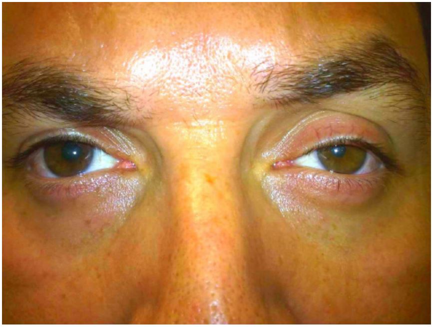
Figure 1: Left blepharoptosis, ipsilateral miosis, and left facial anhidrosis.
References
- Kanagalingam S, Miller NR. Horner syndrome: clinical perspectives. Eye and Brain. 2015; 7: 35-46
- Martin TJ. Horner Syndrome: A Clinical Review. ACS Chem. Neurosci. 2018; 9: 177-186.
- Fernandes-Marques M, Henrique-Barros L, Lopes-Correia B, Lopes da Silva E, Rodrigues-Pinto R. Horner Syndrome After Anterior Revision Surgery for Cervical Spondylotic Myelopathy: A Very Rare Complication. JBJS Case Connect. 2018; 8: 94.
