
Open Access, Volume 9
Hibernoma mimicking a lipoma: A case report of a rare benign tumor
Fatima Buti*; Suhaila Ahmed; Ayesha Amal; Anaum Kabeer; Hadiel Kaiyasah
General Surgery Department, Rashid Hospital, Dubai Academic Health Corporation, Dubai, UAE.
Fatima Buti
General Surgery Department, Rashid Hospital, Dubai Academic Health Corporation, Dubai, UAE.
Email: fatima.buti90@gmail.com
Received : Oct 23, 2023,
Accepted : Nov 21, 2023
Published : Nov 30, 2023,
Archived : www.jclinmedcasereports.com
Abstract
Hibernomas are rare, benign soft tissue tumors developed from fetal brown fat [1]. They account for around 1.6% of benign lipomatous tumors and present as progressive, slow-growing, painless subcutaneous masses [2]. Hibernomas resemble lipomas in their clinical behavior, but their unique imaging and histopathologic features may aid in differentiating them from each other [1]. We report a case of a 19-year-old Emirati woman who presented with painful lower back swelling for one year. The lesion was excised, and the histopathology revealed a diagnosis of hibernoma. The patient had an uneventful postoperative course and was discharged on the second postoperative day. The management approach of hibernomas varies as per the symptoms, our patient was managed with surgical intervention whereas asymptomatic patients can be observed. Surgical resection is the definitive treatment. No local recurrence has been reported.
Keywords: Hibernoma; Benign tumor; Fetal brown tissue; Lipoma.
Copy right Statement: Content published in the journal follows Creative Commons Attribution License (http://creativecommons.org/licenses/by/4.0). © Buti F (2023)
Journal: Open Journal of Clinical and Medical Case Reports is an international, open access, peer reviewed Journal mainly focused exclusively on the medical and clinical case reports.
Citation: Buti F, Ahmed S, Amal A, Kabeer A, Kaiyasah H. Hibernoma mimicking a lipoma: A case report of a rare benign tumor. Open J Clin Med Case Rep. 2023; 2160.
Introduction
Hibernomas are rare benign neoplasms that develop from fetal brown fat. They were first described under the name ‘pseudolipoma’ by the German physician H. Merkel in 1906 and the term hibernoma was proposed by the French anatomist Louis Gery in 1914 because of its resemblance to brown fat in hibernating animals [3,4]. It is prevalent between 30 and 40 years, with a slight male predominance. Hibernoma cells are small, rounded, with numerous cytoplasmic vacuoles or granular cytoplasm surrounding a central nucleus [5]. The etiology of hibernomas is unknown; however, molecular genetics shows chromosome 11q13 translocations, which encode the tumor suppressor genes MEN 1 and AIP [1,3].
Hibernomas are well-circumscribed tumors on computed tomography (CT) and magnetic resonance imaging (MRI), frequently found in subcutaneous tissue, skeletal muscle, or intermuscular fascial planes. The thigh, trunk, upper extremities, head, and neck are common presentation sites. The lipoma-like variant is easily misdiagnosed as atypical lipoma or well-differentiated liposarcoma because of their similar clinical presentation [4]. Hibernomas are benign tumors without risk of malignant transformation or metastases [1]. Asymptomatic hibernoma can be treated conservatively. Incomplete resection increases the likelihood of recurrence, whereas total resection resolves symptomatic hibernomas.
Case Presentation
A 19-year-old Emirati female, a known case of disc prolapse, presented complaining of lower back swelling for one year. On examination, the patient looked alert, and oriented with a BMI of 20.4, and was hemodynamically stable. Local examination revealed a soft, non-tender 4x5 cm lower back lump above the natal cleft. Routine blood investigations were within normal range; targeted ultrasound suggested benign morphology soft tissue fibromuscular lipoma. MRI revealed an atypical lipoma in the subcutaneous region of the back at the level of L4-15 vertebra with disc herniation at the same level.
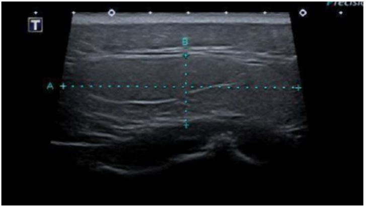
Figure 1: Targeted Ultrasound at L5/S1 level: A well-defined, elliptical-shaped, solid soft tissue lesion measuring approximately 6.5 cm × 1.8 cm situated at the junction of the subcutaneous and muscular plane with no significant internal vascularity, calcifications or cystic component.
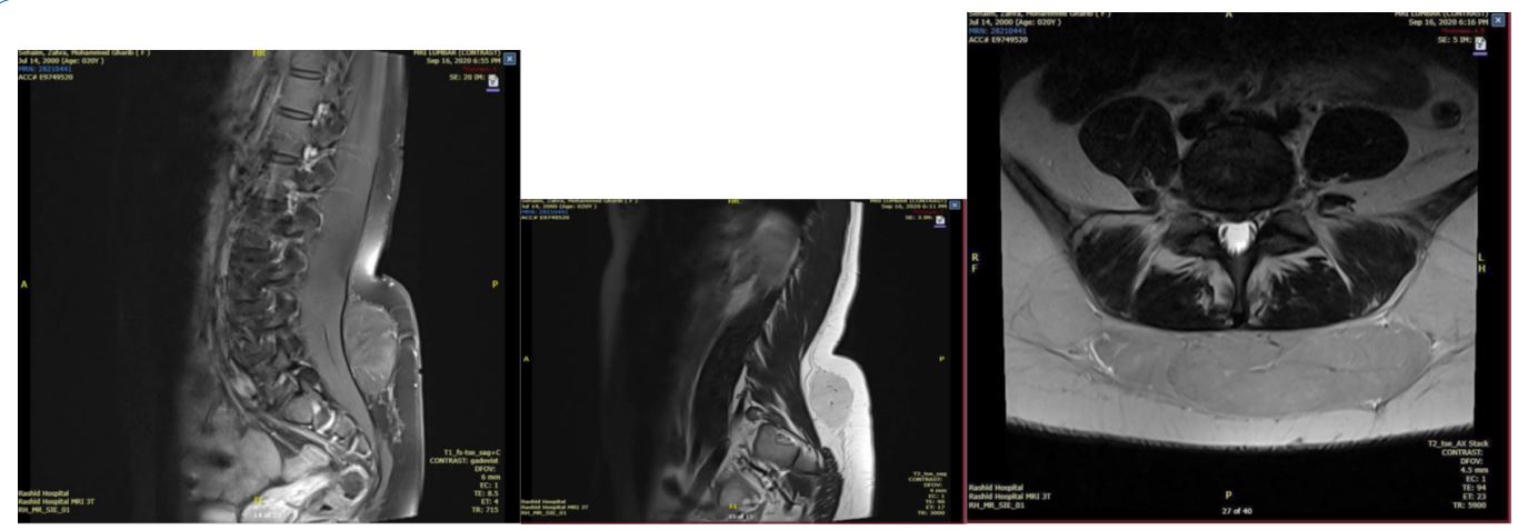
Figure 2: MRI at L5/S1: A large, well-defined T2 hyper-intense lesion measuring about 12 cm in transverse, 3 cm in anteroposterior, and 9 cm at the craniocaudal dimension with thin septa and blood vessels traversing through it, in the subcutaneous region of the back at the level of L4-L5 vertebra with disc herniation at the same level. Under GA, complete surgical
excision was done. The postoperative course was uneventful. The patient was discharged the next day. Histopathology biopsy
revealed: a yellow-colored nodular lobulated mass weighing 145.0 gm and measuring 11.5 x 9.4 x 3.2 cm. Slicing revealed
lobules of adipose tissue.
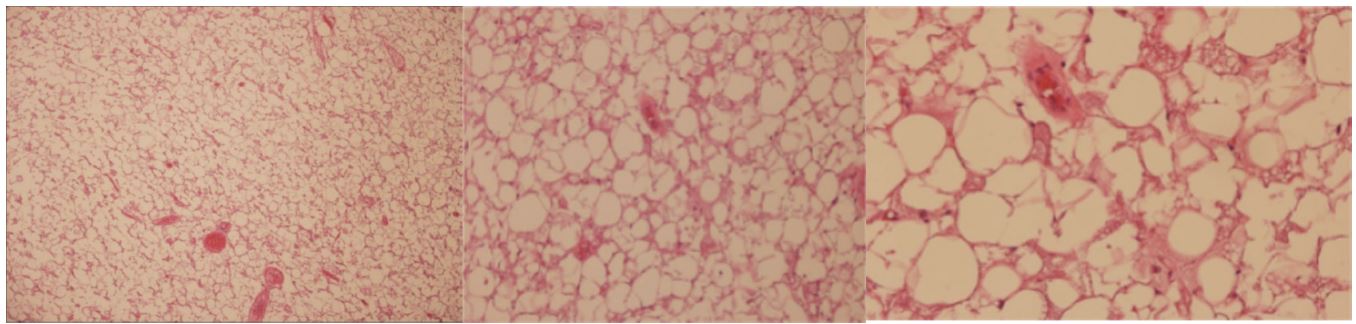
Figure 3: Histopathology section: A benign neoplasm, showing lobules composed of an admixture of palely staining uni
vacuolated fat cells and eosinophilic multivacuolated hibernoma cells with a markedly enlarged cytoplasm filled with course
vacuoles and eosinophilic granules.
In conclusion, the histomorphological features of the specimen were consistent with hibernoma. The patient had an uneventful recovery after the lipoma excision.
Discussion
Hibernoma is a rare, benign, well-circumscribed, slowly growing and well-vascularized soft-tissue tumour arising from the vestiges of fetal brown fat, usually resembling the special fat deposits of hibernating animals. Hibernomas constitute approximately only 1.6% of all benign lipomatous tumours. The tumour depicts a slight male predominance in the third to fourth decade of life, making our case an atypical presentation. The first case report of this tumour appeared in literature in 1906 [4]. There have been less than 200 cases published so far [4]. To the best of our knowledge, up till now there has been no similar case reports in the UAE. As hibernoma is a remnant tissue of brown fat, the thigh is the most frequent location. However, it can appear in other areas (e.g. areas of residual brown fat such as the neck, axilla, back and mediastinum, less frequently the trunk or retroperitoneum). They usually measure 5 to 10 cm in diameter but may reach up to 20 cm [4]. There are no known risk factors for hibernoma [6]. We report the case of a hibernoma in the subcutaneous region of the back at the level of the L4-L5 vertebra. In our case, MRI imaging showed an uncertain appearance; an atypical lipoma was suspected; therefore, we performed an excisional biopsy. The histopathological study confirmed the diagnosis of hibernoma. The differential diagnosis of lipomatous soft-tissue tumors is broad and comprises benign (e.g. lipoma, hibernoma, hemangioma, angiolipoma) and malignant (e.g. liposarcoma) lesions [5]. Despite the use of multimodal imaging techniques, it was highly challenging to radiologically diagnose hibernomas and distinguish them from well-differentiated liposarcomas or atypical lipomas.
Conclusion
Hibernomas should be kept in the differentials when dealing with an atypical lipoma. Conservative management is preferred for asymptomatic hibernomas. Whereas complete surgical resection is the cure for symptomatic patients.
Acknowledgments: We would like to thank Dr. Tasnim Keloth for providing the pathology report for this case.
References
- Daubner D, Spieth S, Pablik J, Zöphel K, Paulus T, Laniado M. Hibernoma – two patients with a rare lipoid soft-tissue tumour. BMC Medical Imaging. BioMed Central. 2015.
- Hibernoma DermNet. (n.d.). Hibernoma | DermNet.
- Feger J. Hibernoma: Radiology Reference Article. Radiopaedia Blog RSS.
- Gavriilidis P, Panselinas G, Zafiriou G. Rare disease: Hibernoma of the thigh: a lipoma-like variant rare tumour mimicking soft tissue sarcoma. PubMed Central (PMC). 2012.
- Hibernoma - An overview | ScienceDirect Topics. (n.d.). Hibernoma – an Overview | ScienceDirect Topics.
- H Reichel, T Rueckl, K Fenwick, A Vogt, N Rudert M, Plumhoff, P. Hibernoma of the Upper Extremity: Complete Case of a Rare but Benign Soft Tissue Tumor. 2019.
