Open Access, Volume 9
A rare case of cutaneous angiosarcoma on the face in an 84-year-old patient: A case report and review of the literature
Sektaoui Soukaina*; Mehsas Zoubida; Meziane Mariame; Nadia ismaili; Benzekri Leila; Senouci Karima
Department of Dermatology & Venereology, Ibn Sina University Hospital, Faculty of Medicine and Pharmacy of rabat, Mohamed V University, Rabat, Morocco.
Sektaoui Soukaina
IDepartment of Dermatology & Venereology, Ibn Sina University Hospital, Faculty of Medicine and Pharmacy of Rabat, Mohamed V University, Rabat, Morocco.
Email: Soukaina.sektaoui@gmail.com
Received : January 27, 2023,
Accepted : February 21, 2023
Published : February 28, 2023,
Archived : www.jclinmedcasereports.com
Abstract
Cutaneous angiosarcoma is a rare and aggressive form of cancer that affects the blood vessels and lymph vessels of the skin and soft tissues. It is estimated to account for only 2% of all skin malignancies, and its incidence can vary depending on the location and organ affected. In this case report, we present a unique case of an 84-year-old woman with a history of traditional tattoos on her chin, nose, and neck more than 50 years ago who developed an angiosarcoma on her left cheek. The patient underwent non-oncological surgical excision and adjuvant radiotherapy. The pathological examination showed a low-grade differentiated angiosarcoma. The aim of this case report is to raise awareness of the rarity and aggressiveness of angiosarcoma, especially on the face, and the importance of early diagnosis and treatment.
Keywords: Angiosarcoma; Cutaneous; Face; Traditional tattoos; Case report
Copy right Statement: Content published in the journal follows Creative Commons Attribution License (http://creativecommons.org/licenses/by/4.0). © Soukaina S (2023)
Journal: Open Journal of Clinical and Medical Case Reports is an international, open access, peer reviewed Journal mainly focused exclusively on the medical and clinical case reports.
Citation: Soukaina S, Zoubida M, Mariame M, Ismaili N, Leila B, et al. A rare case of cutaneous angiosarcoma on the face in an 84-year-old patient: A case report and review of the literature. Open J Clin Med Case Rep. 2023; 1985.
Introduction
Cutaneous angiosarcoma is a rare and aggressive form of cancer that affects the blood vessels and lymph vessels of the skin and soft tissues. It is estimated to account for only 2% of all skin malignancies, and its incidence can vary depending on the location and organ affected. It commonly affects the head and neck region, specifically the scalp, face and neck [1]. The malignant cells of angiosarcoma form within the walls of blood vessels and lymph vessels, leading to the formation of tumors that can grow quickly and invade surrounding tissue [2]. In this case, we present the case of a 84-year-old woman with an angiosarcoma on her left cheek. This case is unique as it’s developed on the skin of the face, and also, the patient’s history of traditional tattoos on her chin, nose, and neck more than 50 years ago.
Case Presentation
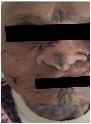
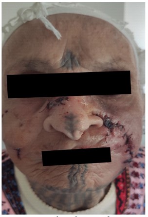
This is a 84-year-old patient who has been being treated for ischemic heart disease for 10 years. She had traditional tattoos on her chin, nose, and neck more than 50 years ago. For the past 3 months, she has had a painless, bleeding, rapidly growing swelling on her left cheek and a lump in her nasal furrow. Clinical examination found a 7 cm, ulcerated, purple-red, bleeding tumefaction on her left cheek, resting on a red, warm, and painless base, as well as a purple-red lump in her nasal furrow. The biological test results were normal. A skin biopsy was performed and confirmed the diagnosis of a low-grade differentiated angiosarcoma. A facial MRI was also performed and revealed a left cheek process with local invasion and no signs of regional invasion. The thoraco-abdomino-pelvic scan did not find any associated metastases. The patient underwent non-oncological surgical excision (due to advanced local invasion). The postoperative period was uneventful. The tumor sample was sent for anatomic-cyto-pathological analysis and found to have excision with margin limits. The patient received adjuvant radiotherapy as the surgery was non-oncological.
Discussion
Angiosarcoma is a rare and aggressive form of cancer that affects the blood vessels and lymph vessels of the skin and soft tissues. It accounts for only 2% of all skin malignancies, and its incidence can vary depending on the location and organ affected [1]. Previous studies have found that the most aggressive presentation of angiosarcoma is on the face [3]. Lesions typically present as well-demarcated, often traversed by areas of subcutaneous hematoma [4]. They may appear as bluish macules, similar to bruising, and may have a peripheral erythematous ring, satellite nodules, intratumoral hemorrhage, and a tendency to bleed spontaneously or with minimal trauma [5]. In this case, the patient presented with sudden onset of ulcerative lesions on the left cheek that appeared 3 months prior to surgery. Examination revealed a 7 cm, ulcerated, purple-red, bleeding tumefaction, resting on a red, warm, and painless base, as well as a purple-red lump in her nasal furrow. Lymph node involvement in the neck was not found. Pathological examination was conducted to establish the diagnosis [6]. Morphologically, cutaneous angiosarcoma is similar to other tumors, showing infiltrative growth of irregular vascular spaces that anastomose with characteristic dissection between collagen bundles [7]. The tumor can be classified into two grades: low-grade lesions with irregular vascular channels lined with atypical endothelial cells, and high-grade lesions that are sheets of undifferentiated pleomorphic cells, often difficult to distinguish from carcinoma [1]. Biopsy and immuno-histochemistry are used to confirm the diagnosis. Treatment options include wide surgical resection, which may be combined with free muscle flaps and skin grafts, and may be followed by radiotherapy and/or chemotherapy [8]. Some studies have shown that a combination of radical surgery and adjuvant radiation therapy is optimal [9]. In advanced local invasion cases like ours, a non-oncological surgical excision was performed, followed by adjuvant radiotherapy. The 5-year survival rate in the head and neck region varies widely and tends to be low [10].
Conclusion
Cutaneous angiosarcoma is a rare and aggressive form of cancer that affects the blood vessels and lymph vessels of the skin and soft tissues. This case report presents a unique case of an 84-year-old woman with a history of traditional tattoos on her chin, nose, and neck more than 50 years ago who developed an angiosarcoma on her left cheek. The patient underwent non-oncological surgical excision and adjuvant radiotherapy. The pathological examination showed a low-grade differentiated angiosarcoma. The importance of early diagnosis and treatment of angiosarcoma, especially on the face, should be emphasized to increase awareness and improve outcomes for patients.
Acknowledgements: All the authors provide valuable imput and feedback on the research and writing of the article, there was no funding resources and no conflict of interest.
References
- Andersen NJ, Froman RE, Kitchell BE, Duesbery NS. Soft Tissue Tumors. Clinical and molecular biology of angiosarcoma. 2011.
- James W, Elston D, Treat J, Rosenbach M, Micheletti R. In: Andrews’ Dis. Ski. 13th ed. James W., Elston D., Treat J., Rosenbach M., Micheletti R., editors. Dermal and subcutaneous tumours, Elsevier; Philadelphia: 2019.
- Bernstein J, Irish JC, Brown D, Goldstein D, Chung P, et al. Survival outcomes for cutaneous angiosarcoma of the scalp versus face. Head Neck. 2017.
- Kim JJ, Seo S, Kim JE, Jung SH, Song SY, et al. Clinical outcomes of angiosarcoma: a single institution experience. Cancer Commun. 2019; 39: 4-7.
- Zhang Y, Yan Y, Zhu M, Chen C, Lu N, et al. Clinical outcomes in primary scalp angiosarcoma. Oncol Lett. 2019; 18: 5091-5096.
- Manaster BJ. Soft-tissue Masses: Optimal Imaging Protocol and Reporting. 2013; 201: 505-514.
- Calonje JE. Fifth edit. Elsevier Inc.; 2020. Tumors of Blood Vessels and Lymphatics.
- Miqueloti MB, Gonzaga CB, Ribeiro V.da S.A, Filho E.Glória, Freire RAV. Scalp reconstruction after wide resection of an angiosarcoma. Rev Bras Cir Plástica Braz J Plast Surg. 2019; 34.
- Oashi K, Namikawa K, Tsutsumida A, Takahashi A, Itami J, et al. Surgery with curative intent is associated with prolonged survival in patients with cutaneous angiosarcoma of the scalp and face -a retrospective study of 38 untreated cases in the Japanese population. Eur J Surg Oncol. 2018; 44: 823-829.
- Patel SH, Hayden RE, Hinni ML, Wong WW, Foote RL, et al. Angiosarcoma of the scalp and face: theMayo clinic experience. JAMA Otolaryngol. 2015; 141: 335-340.


