
Open Access, Volume 11
Challenging the limits of myomectomy during pregnancy for improved fetal maternal outcomes: A case series
Seema Mehrotra1; Shyam Pyari Jaiswar2*; PushpLata Sankhwar1; Manju Lata Verma1; Shruti Gupta1; Sneha Agarwal1; Ummehani Sidiqui1
1Professor, Department of Obstetrics and Gynaecology, King George’s Medical University, India.
2Professor & Head, Department of Obstetrics and Gynaecology, Era Medical University, India.
Shyam Pyari Jaiswar
Professor & Head, Department of Obstetrics and Gynaecology, Era Medical University, Lucknow 226003, Uttar Pradesh, India.
Tel: +91-9415023358;
Email: spjaiswar59@gmail.com
Received : February 14, 2025,
Accepted : March 10, 2025
Published : March 28, 2025,
Archived : www.jclinmedcasereports.com
Abstract
Introduction: Uterine myomas afflict 2-10% of pregnant women. Myomas complicate the course in every ten patients, leading to poor pregnancy outcomes such as placental abruption, early rupture of membranes, placenta previa, preterm labor, fetal malpresentation. Myomectomy is rarely performed during pregnancy due to the risk of miscarriage and uncontrollable bleeding. Surgical therapy is required in some cases where conservative management fails to control symptoms and there is a significant risk of negative pregnancy outcomes. This article explores the safety, dangers, and benefits of prenatal myomectomy.
Methodology: This is a retrospective study of six individuals who had myomectomy between 16- and 19-weeks gestation at our tertiary care center. The decision for surgical management was based on large fibroid size & refractory pain. Myoma was enucleated via transverse incision & reconstruction of uterine wall was done in double layer.
Results: None of the patients in our study had miscarriage, preterm labour, premature rupture of membranes, placental abruption. 5 patients underwent elective caesarean sections, 1 patient underwent vaginal delivery following labor induction, and all patients delivered at term with a median gestational age of 37 weeks. None of the patients delivered a growth-restricted fetus or a fetus with a congenital anomaly. No adverse maternal or fetal outcome was reported.
Conclusion: Antenatal myomectomy should not be done as a routine procedure. However, when performed on carefully chosen patients under competent supervision, it is a safe operation that produces positive pregnancy results without the usual difficulties associated with myoma presence.
Keywords: Antenatal myomectomy; Caesarean section; Myoma; Pregnancy complications; Uterine fibroid.
Abbreviations: ASD: Atrial Septal Defect; BMI: Body Mass Index; IUGR: Intrauterine Growth Restriction; LSCS: Lower Segment Cesarean Section; MRI: Magnetic Resonance Imaging; PAH: Pulmonary Arterial Hypertension; PRBC: Packed Red Blood Cells; PROM: Premature Rupture of Membranes; USG: Ultrasonography.
Copy right Statement: Content published in the journal follows Creative Commons Attribution License (http://creativecommons.org/licenses/by/4.0). © Jaiswar SP (2025)
Journal: Open Journal of Clinical and Medical Case Reports is an international, open access, peer reviewed Journal mainly focused exclusively on the medical and clinical case reports.
Citation: Mehrotra S, Jaiswar SP, Sankhwar P, Verma ML, Gupta S, et al. Challenging the limits of myomectomy during pregnancy for improved fetal maternal outcomes: A case series. Open J Clin Med Case Rep. 2025; 2333.
Introduction
Uterine fibroids are the most frequent benign smooth muscle tumors, accounting for 2-10% of cases during pregnancy. Smooth muscle cells and fibroblasts produce extracellular matrix [1]. Although most myomas are asymptomatic, problems can arise in one out of every ten women during the prenatal period [2,3]. The increased placental hormones and uterine blood flow during pregnancy cause an increase in fibroid volume, which may complicate the pregnancy. The most common consequences are abdominal pain, fever, and vaginal bleeding. The frequency of pain rises with growth, particularly for fibroids larger than 5 cm in diameter [3,4]. Uterine fibroids can raise the risk of miscarriage, fetal growth limitation, fetal malpresentation, placental abruption, premature membrane rupture, placenta previa, preterm labor, and cesarean birth. Furthermore, uterine fibroids can cause difficulties during labor and delivery, including aberrant uterine contractile activity, fetal discomfort, uterine atony, and postpartum hemorrhage [1]. Overall, the management of severe symptoms & signs caused by uterine fibroids during pregnancy poses a challenge, because pharmacological approaches for alleviating pain during pregnancy are limited [1]. In selected cases, when other treatment strategies fail to manage symptoms or there is a huge myoma filling the abdominal cavity and restricting the growth of the fetus and there is a substantial risk of adverse pregnancy outcomes, a surgical approach during pregnancy might be considered. Myomectomy is usually not advocated during pregnancy because of possible complications such as severe haemorrhage, uterine rupture, miscarriage and preterm labor [5,6]. However, complication can be prevented in a well optimized surgery by a skilled surgeon, with comprehensive peri-operative preparation and counselling.
Material and Methods
This study was conducted as a retrospective single centre case series of 6 patients. Records of patients who underwent antenatal myomectomy at our tertiary care centre between January 2023 to March 2024 were collected. Informed consent was taken from all the patients. The antenatal course, clinical characteristics, perioperative management and pregnancy outcome are discussed. The following criteria was used for myomectomy in our study-(i) gestational age >14 weeks; (ii)large(>15 cms) size of uterine fibroid that prevented proper continuation of pregnancy; (iii) symptomatic uterine fibroids(severe abdominal pain, rapid growth) (iv) a distance between the leiomyoma and the endometrial cavity >5 mm, in order to avoid opening of the endometrial cavity; and (iv) the provision of a signed consent form, after the patients had been informed of the risks of surgical intervention [1,7].
Surgical approach was deferred in patients fulfilling following criteria: patients who refused surgery or did not sign informed consent; presence of chromosomal abnormalities and/or congenital malformations of the fetus; and absence of urgent indications for myomectomy during pregnancy [1].
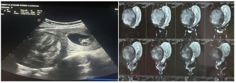
Figure 1: Ultrasonography and MRI film showing large subserosal uterine fibroid in relation to fetus.
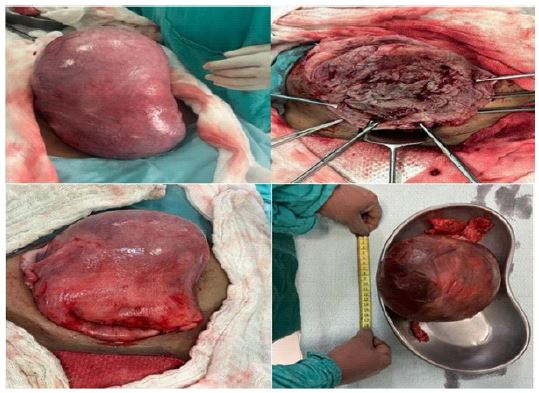
Figure 2: Perop image during myomectomy showing subserosal myoma, base of myoma after enucleation, uterine wall after reconstruction & gross image of removed myoma.
All patients were admitted to hospital between 10-16 weeks of gestation for clinical evaluation, laboratory workup, transvaginal & abdominal ultrasonography and electrocardiography. Fibroid characteristics such as size, location, distance from the lower pole of the myoma to the uterine cavity, and presence of areas degeneration were studied (Figure 1). Doppler ultrasonography & MRI was further used to study vascularsupply and mapping of leiomyoma, when needed. Blood typing and cross matching was done. One unit of blood was readied as a precautionary measure to mitigate the risks associated with significant blood loss during surgery. Laparotomy was performed via midline vertical incision that extended over the umbilicus under regional anaesthesia (or endotracheal anaesthesia when needed), after ruling out congenital malformations & chromosomal anomalies. Myoma was enucleated via transverse incision given over fibroid capsule followed by blunt and sharp dissection. Following myomectomy reconstruction of uterine wall was done in double layer via polyglactin no.1-0 suture material (Figure 2). Haemostasis was achieved via multiple sutures at myoma base. In postoperative period tocolytics were given to prevent uterine contrac tions. Intravenous Isoxsuprine Hydrochloride drip was started intraoperatively and continued for 48 hours which was then tapered and patient put on oral medication for 1 week. Progesterone support in form of Hydroxyprogesterone Hexanoate 500 mg given intramuscularly before surgery and every week till 36 weeks to prevent possible miscarriage and preterm labour. All patients were given antibiotics and analgesics for 5 days postoperatively. Fetal well being was checked by ultrasonography at the end of surgery. Removed myoma was sent for histopathological examination. After discharge patient were closely followed up every 2 weekly for antenatal surveillance on OPD basis. Ultrasonography was performed monthly to monitor fetal growth. Maternal and fetal outcomes & complications were studied.
Results
Clinical and surgical characteristics of antenatal myomectomy patients are discussed in (Table 1).
• Demographic profile - All cases that presented to us were primigravida of Asian ethnicity with mean age of 30 years & BMI of 25.6 kg/m2.
• Medical history & symptomatology - 5 cases had uneventful previous medical history and 1 case had acyanotic congenital heart disease (ASD with mild MR with moderate PAH). There was persistent abdominal pain not responding to conservative management associated with vomiting & epigastric discomfort in all cases. Two of the cases presented with rapid growth of leiomyoma in pregnancy. Patients were hospitalized after onset of symptoms. On average, uterine fibroid related symptom onset occurred at 13.6 weeks. There was no history of oral or injectable contraceptive use.
• Management - Laparotomy was performed at the mean gestational age of 17.8 weeks (16-19 weeks) with removal of single large fibroid having mean size and weight of and 2.86 kg respectively. Among removed fibroids 2 were intramural (FIGO IV), 3 were subserous (FIGO V & VI) and 1 was subserous pedunculated fibroid with torsion (FIGO VII). 50% of cases were done under spinal plus epidural anaesthesia and rest 50% under epidural plus endotracheal anaesthesia.
• Surgical variables - Mean duration of surgery was 80 mins and average blood loss was 460 cc with most of the patients requiring 1-unit PRBC transfusion.
• Histology - On histopathology 4 myomas revealed hyaline degeneration,1 red degeneration and 1 necrosis.
• Postoperative period - Mean duration of hospital stay was 8 days without any postoperative complications.
• Maternal & fetal outcome - None of the patient had spontaneous miscarriage or went into preterm labour. All our patients delivered at term gestation, 5 via lower segment caesarean section and 1 via vaginal delivery after induction of labour. All were live births with median fetal weight and APGAR score of 3.0 kg and 6/8 respectively. None of the infants had gross structural malformation. Postpartum period was uneventful in all women.
Table 1: Clinical and surgical characterstics of patients.
| Variable | Case 1 | Case 2 | Case 3 | Case 4 | Case 5 | Case 6 | Mean |
|---|---|---|---|---|---|---|---|
| Age | 32 | 25 | 27 | 32 | 28 | 35 | 30 |
| BMI (kg/m2) | 24.6 | 27.2 | 26 | 25.4 | 26.1 | 24.8 | 25.6 |
| Parity | Primigravida | Primigravida | Primigravida | Primigravida | Primigravida | Primigravida | - |
| Symptoms | Abdominal pain | Abdominal pain | Abdominal pain | Abdominal pain | Abdominal pain | Abdominal pain | |
| Rapid growth | Rapid growth | ||||||
| Gestational age atsymptom onset | 16 weeks | 13 weeks | 10 weeks | 13 weeks | 15 weeks | 15 weeks | 13.5weeks |
| Medical History | None | None | None | Congenital heartdisease | None | None | |
| Gestational age atmyomectomy | 19 weeks | 19 weeks | 16 weeks | 17 weeks | 18 weeks | 18 weeks | 18 weeks |
| Fundal heighton abdominalexamination | 34 weeks | 34 weeks | 32 weeks | 32 weeks | 32 weeks | 34 weeks | |
| Anaesthesia during myomectomy | SA+EA | GA +EA | SA+EA | GA+EA | SA+EA | GA +EA | |
| Gross size of myoma | 19×15×12 cm | 18×15×16 cm | 18×14×12 cm | 15×12×10 cm | 15×10×14 cm | 17×14×12 cm | 16×13×13 cm |
| Weight of myoma | 2.85 kg | 3.7 kg | 3.0 kg | 2.4 kg | 2.5 kg | 2.75 kg | 2.86 kg |
| Type of Myoma | Subserous | Subserous | Intramural | Subserous | Intramural | Subserous pedunculated | |
| FIGO stage | VI | V | IV | V | IV | VII | |
| Blood loss duringsurgery | 400 cc | 500 cc | 500 cc | 450 cc | 550 cc | 400 cc | 460 cc |
| Duration of surgery | 70 mins | 90 mins | 90 mins | 80 mins | 80 mins | 60 mins | 80 mins |
| PRBC transfusion | 1 unit | 1 unit | 1 unit | 1 unit | 1 unit | None | - |
| Hospital Stay | 8 days | 8 days | 8 days | 9 days | 10 days | 6 days | 8 days |
| ICU stay | 0 | 0 | 0 | 1 day | 0 | 0 | |
| Mode ofdelivery | LSCS | LSCS | LSCS | LSCS | LSCS | Vaginal | - |
| Gestational age atdelivery | 37 weeks | 37 week2 day | 38 week | 37 weeks | 37 weeks | 39 weeks | 37.5weeks |
| Fetal Outcome | Fullterm live birth | Fullterm live birth | Fullterm live birth | Fullterm live birth | Fullterm live birth | Full termlive birth | |
| Fetal weight/APGAR scores | 2.9 kg(6/8) | 2.96 kg(7/8) | 2.8 kg(7/8) | 3.2 kg(6/8) | 3.0 kg(6/8) | 3.5 kg(6/7) |
Discussion
To the best of our knowledge, this is the first case series from India that attempted to analyze myomectomy during pregnancy. Uterine fibroids affect 2-10% of pregnant women [2]. The current tendency to delay the age of pregnancy and the increased number of mothers over the age of 30 years have resulted in a significant increase in the frequency of pregnant women with uterine fibroids in recent years [1]. Approximately 30% of pregnancies with uterine fibroids suffer from obstetric complications. The performance of myomectomy during pregnancy is very much debated. The symptoms of uterine fibroids in pregnancy often respond to medical therapy, but in selected cases, when conservative management fails or there is a huge myoma & there is substantial risk of adverse pregnancy outcomes, a surgical approach might be necessary. The most commonindications for myomectomy reported in the literature [2,5,8] are: acute pelvic pain unresponsive to medical therapy of >72 h, rapid growth in myoma, mass compressing the pelvic organs (leading to urinary retention, constipation), and a high risk of fetal adverse events (fetal compression syndrome, oligohydramnios, intrauterine growth restriction, hemorrhages, and placental site abnormalities).
However, myomectomy in pregnancy is associated with increased the risk of bleeding complications & adverse pregnancy outcomes such as miscarriage, maternal and fetal infections, preterm delivery, and uterine rupture [1]. However, in some cases, as discussed earlier, surgical removal of myoma is the only choice to improve patient outcome.
'Leiomyomas in pregnancy are associated with spontaneous abortion rates of as high as 20% [9]. The suggested mechanism is compressed endometrial vascular supply, that adversely affects the fetus resulting in abortion. In contrast to this, none of the patients in our study had miscarriage or required hysterectomy. This shows that antenatal myomectomy is effective in preventing this complication.
In our study no patient had preterm labor and premature rupture of membranes. This shows that elective surgery decreases the incidence of preterm delivery in accordance with the results described by Handa et al. [9].
Studies have shown a 3-fold increase in occurrences of abruptio placentae in pregnant women with uterine fibroids [10]. In our study, no case of abruptio placentae occurred, demonstrating that myomectomy is effective in preventing this complication.
Large fibroids can cause spatial restriction, potentially leading to hindered fetal growth and limb defects [11]. In our study, none of the patients delivered a growth-restricted fetus or a fetus with a congenital anomaly. This demonstrates that performing antenatal myomectomy improves neonatal outcome.
This shows that patients with untreated symptomatic uterine fibroids during pregnancy seem to have a worse pregnancy outcome than patients treated surgically. Despite these findings, laparotomic myomectomy should be considered in pregnancy only when there are appropriate indications.
In our study antenatal myomectomy was performed at the mean gestational age of 17.8 weeks after ruling out congenital malformations & chromosomal anomalies. Epidural anaesthesia was given in all cases as it provides effective and long-term anaesthesia during surgery and in the postoperative period, along with spinal and endotracheal anaesthesia, where needed. The most common complication of myomectomy during gestation is massive bleeding often requiring multiple blood transfusions. This presents a surgical challenge as tourniquets and local injections of vasoconstrictive agents are avoided since it could reduce uterine blood supply [13]. In our study haemostasis was achieved by using multiple haemostatic clamps and sutures at the base of myoma &uterine scar was closed in two layers using polyglactin1-0 suture. One-unit PRBC transfusion was required postoperatively. In our study most of the fibroids were subserous and intramural located at uterine fundus. The lack of patients with submucosal fibroids may be due to the fact that previously identified submucosal leiomyomas were removed prior to pregnancy as these tend to be symptomatic and may contribute to failure to conceive and pregnancy loss.
Histopathology of the removed myomas revealed hyaline degeneration (4 cases), red cell degeneration (1 case) and necrosis (1 case). Degenerative changes might be due to rapid growth or torsion of the myoma leads to deficiency in blood supply, with secondary tumor anoxia and necrosis [3]. All patients delivered at term with median gestational age of 37 weeks, mostly by elective caesarean section except one patient delivered vaginally who had pedunculated subserosal myoma. No adverse maternal or fetal outcome was reported.
Conclusion
Our study shows that antenatal myomectomy, when performed in carefully selected patients, can improve pregnancy outcomes and help prevent complications related to fibroids. However, it should not be recommended as a routine procedure. In cases where fibroids cause significant complications, such as refractory pain or pose risks to the pregnancy due to their large size, antenatal myomectomy may offer an opportunity to improve both maternal and fetal health.
The decision to proceed should be individualized, taking into account the size of the fibroid, associated symptoms, and the overall clinical context, and the surgeon’s expertise.
Declarations
Ethical approval: The study was conducted in accordance with the ethical standards of the Institutional Ethical Clearance Committee for Human Research at King George’s Medical University, Lucknow, Uttar Pradesh, India. A waiver for ethical clearance was obtained on 07/06/2024. The waiver document is attached as a supplementary file.
Informed consent: Written informed consent was obtained from all patients prior to participation in the study, including consent for publication of anonymized data.
Funding: This study was not funded by any individual or agency.
Conflicts of interest: The authors declare no conflicts of interest.
Data availability: The datasets used and analyzed during the current study are available from the corresponding author upon reasonable request.
Acknowledgments: The authors sincerely acknowledge the unwavering support of colleagues, nursing staff, and institutional authorities for their assistance in conducting this study.
References
- Babunashvili EL, Son DY, Buyanova SN, Schukina NA, Popov AA, Chechneva MA, et al. Outcomes of Laparotomic Myomectomy during Pregnancy for Symptomatic Uterine Fibroids: A Prospective Cohort Study. J Clin Med. 2023; 12: 6406.
- Saccardi C, Visentin S, Noventa M, Cosmi E, Litta P, Gizzo S. Uncertainties about laparoscopic myomectomy during pregnancy: a lack of evidence or an inherited misconception? A critical literature review starting from a peculiar case. Minim Invasive Ther Allied Technol. 2015; 24: 189-94.
- Spyropoulou K, Kosmas I, Tsakiridis I, Mamopoulos A, Kalogiannidis I, Athanasiadis A, et al. Myomectomy during pregnancy: A systematic review. Eur J Obstet Gynecol Reprod Biol. 2020; 254: 15-24.
- Wang HM, Tian YC, Xue ZF, Zhang Y, Dai YM. Associations between uterine fibroids and obstetric outcomes in twin pregnancies. International Journal of Gynecology& Obstetrics. 2016; 135: 22-27.
- Allameh Z, Allameh T. Successful Myomectomy in the Second Trimester of Pregnancy. Adv Biomed Res. 2019; 8: 60.
- Loverro Giuseppe, Damiani Gianluca Raffaello, Malvasi Antonio, Loverro Matteo, Schönauer Luca, et al. Myomectomy during pregnancy: an obstetric overview and two case-series. Minerva obstetrics and gynecology. 2021; 73.
- Vitale Salvatore Giovanni, Padula Francesco, Gulino Ferdinand. Management of uterine fibroids in pregnancy. Current opinion in obstetrics &gynecology. 2015; 27.
- Dinglas C, Kunzier N, Sanchi J, et al. Ultrasound-guided manipulation of fetal entrapment by a large uterine fibroid. Am J Obstet Gynecol. 2015; 213: 870.e1-2.
- Handa N, Anjali. Feto maternal outcomes of fibroid in pregnancy: a retrospective observational study. Int J Reprod Contracept ObstetGynecol. 2023; 12: 3080-5.
- Tîrnovanu MC, Lozneanu L, Tîrnovanu ŞD, Tîrnovanu VG, Onofriescu M, Ungureanu C, Toma BF, Cojocaru E. Uterine Fibroids and Pregnancy: A Review of the Challenges from a Romanian Tertiary Level Institution. Healthcare (Basel). 2022; 10: 855.
- Mihaela Camelia Tîrnovanu, Ludmila Lozneanu, Ştefan Dragoş Tîrnovanu, Vlad Gabriel Tîrnovanu, Mircea Onofriescu, Carmen Ungureanu, et al. Uterine Fibroids and Pregnancy: A Review of the Challenges from a Romanian Tertiary Level Institution, Healthcare. 2022; 10: 5.
- Algara AC, Rodríguez AG, Vázquez AC, Valladares FEC, Ramírez PG, Padilla EL, et al. Laparoscopic Approach for Fibroid Removal at 18 Weeks of Pregnancy. Surg Technol Int. 2015; 27: 195–197.
- Kongnyuy EJ, Wiysonge CS. Interventions to reduce haemorrhage during myomectomy for fibroids. Cochrane Database Syst Rev. 2014; 2014: CD005355.
